
Fig. 2
- ID
- ZDB-FIG-110804-4
- Publication
- Murphy et al., 2011 - A Src-Tks5 Pathway Is Required for Neural Crest Cell Migration during Embryonic Development
- Other Figures
- All Figure Page
- Back to All Figure Page
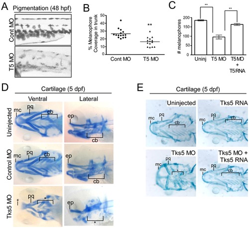
Decreased Tks5 expression results in neural crest-derived defects. (A–B) Melanophores within the trunk region above the yolk sac extension in control MO-injected and Tks5 MO-injected embryos were qualitatively (A) and quantitatively (B) analyzed. n = 15 embryos and SEM is shown by bar. p values obtained from Student′s t-test. ** denotes p<0.01. (C) Melanophores present in the dorsal, ventral, and lateral pigment lines were quantified to determine degree of murine Tks5 rescue of the decreased pigmentation seen in morphants. Mean values (n = 3) and SEM are shown in graph. p values obtained from Student′s t-test. ** denotes p<0.01. (D) Alcian blue staining was performed on indicated embryos to identify craniofacial structures (Meckel′s cartilage (mc), palatoquadrate (pq), ceratobranchials (ch), ethmoid plate (ep)). (*) denotes missing structures. (E) Alcian blue staining was performed on indicated embryos to determine if murine Tks5 could rescue craniofacial defects seen in morphants. Structures were identified as in (D). (*) denotes missing structures. |
| Fish: | |
|---|---|
| Knockdown Reagents: | |
| Observed In: | |
| Stage Range: | Long-pec to Day 5 |