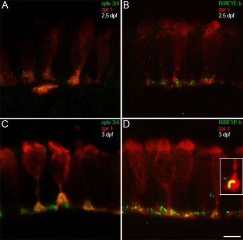
Complexin 3/4 concentrates in retinal photoreceptor terminals concomitant with RIBEYE b. (A) The zpr 1/FRet 43 monoclonal antibody (red) and the pan-complexin 3/4 polyclonal (green) were also used to localize these complexins in developing retinal photoreceptors. At 2.5 dpf, retinal photoreceptors are most prominent in the ventronasal patch, where complexin 3/4 immunoreactivity appears in some photoreceptor terminals. (B) RIBEYE b (green) has also started to cluster in photoreceptor terminals (red) in the outer plexiform layer at 2.5 dpf. (C) At 3 dpf, complexin 3/4 (red) is highly expressed in zpr 1/FRet 43-positive (red) and zpr 1/FRet 43-negative photoreceptor terminals in the ventronasal patch. (D) Pleiomorphic RIBEYE b (green) expression is found at 3 dpf in zpr 1/FRet 43-positive (red) and zpr 1/FRet 43-negative photoreceptor terminals in the ventronasal patch. The inset shows a zpr 1/FRet 43-positive terminal containing curvilinear RIBEYE b immunoreactivity that may correspond to a ribbon. Scale bar = 5 μm.
|
