
Fig. 6
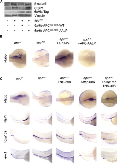
apc Control of Cellular Fate and Differentiation Are Mediated by ctbp1 and Are β-Catenin Independent (A) Protein from 48 hpf apcmcr zebrafish embryos injected with either 6 x His-APC955–2075 or 6 x His-APC955–2075-AALP were subjected to western blot analysis for β-catenin (top), ctbp1 (second), 6 x His (third), or vinculin (bottom). (B) Embryos injected as above were fixed at 72 hpf and subjected to in situ hybridization for i-fabp. (C) apcmcr embryos were treated with DMSO or NS-398 or injected with ctbp1-directed morpholino in the presence or absence of NS-398. At 72 hpf, the embryos were fixed and subjected to in situ hybridization for i-fabp, NaPi, hoxa13a, or evx1. All images are representative of at least three independent experiments. |
| Genes: | |
|---|---|
| Fish: | |
| Knockdown Reagent: | |
| Anatomical Terms: | |
| Stage: | Protruding-mouth |
Reprinted from Cell, 137(4), Phelps, R.A., Chidester, S., Dehghanizadeh, S., Phelps, J., Sandoval, I.T., Rai, K., Broadbent, T., Sarkar, S., Burt, R.W., and Jones, D.A., A two-step model for colon adenoma initiation and progression caused by APC loss, 623-634, Copyright (2009) with permission from Elsevier. Full text @ Cell