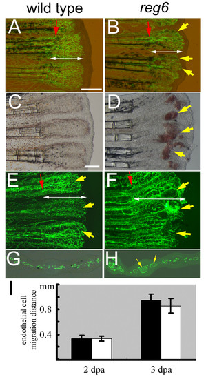
Normal migration of regenerating endothelial cells in zebrafish reg6 mutants. (A, B) Combined bright field and green fluorescent images of wild-type TG(fli1:EGFP)y1 (A) and reg6;TG(fli1:EGFP)y1 (B) regenerating fins at 2 days post amputation (dpa) showing the distance (designated by white double arrows) that the blood vessel endothelial cells (ECs) have migrated. Yellow arrows, dilated vessels of reg6 mutants. (C-F) In 3-dpa regenerates, wild-type fish have formed normal capillary plexuses (C, yellow arrows in E)., but reg6 fish (D, bright field; F, green fluorescent) have developed numerous swollen vessels (yellow arrows in D and F) resulting in blood cell accumulation (red spots in D). (G, H) Cross-sections of 3-dpa regenerates showing normal regenerating vessels in wild-type (G) and dilated vessels in reg6 fish (H, yellow arrows). Red arrows, amputation plane. Scale bars, 300 μm. (I) Quantitative data of the migration distance of regenerating ECs in wild-type (black bars) and reg6 fish (white bars) at 2 and 3 dpa.
|
