
Fig. 6
- ID
- ZDB-FIG-080326-33
- Publication
- Boldajipour et al., 2008 - Control of chemokine-guided cell migration by ligand sequestration
- Other Figures
- All Figure Page
- Back to All Figure Page
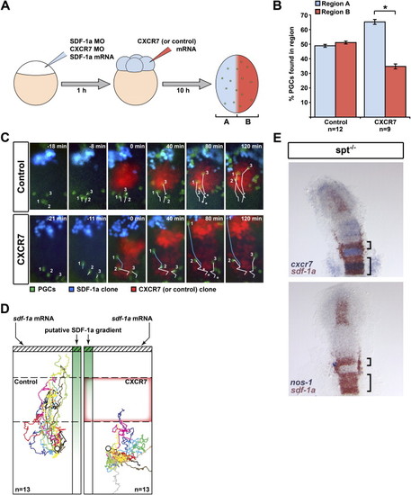
CXCR7 Affects the Direction of Germ Cell Migration In Vivo (A) A schematic representation of the experimental manipulations generating a CXCR7 expression domain (red, Region A) superimposed on uniform SDF-1a expression (blue, Region B). PGCs are depicted in green. (B) In contrast to control experiments, PGCs vacated the CXCR7-expressing B region (p value < 0.001, t test). n signifies the number of embryos examined, and error bars represent SEM. (C) Snapshots of representative time-lapse movies with germ cells (green) migrating toward a transplanted source of SDF-1a (blue) in SDF-1-deficient embryos. A transplant of cells (red) expressing either CXCR7 or control protein was placed at the migration path. In control experiments (upper panel), germ cells (white tracks labeled 1–3) readily traverse the transplant toward the source of SDF-1a. Asterisks denote the starting points. When encountering a CXCR7-expressing transplant (lower panel), the migration toward the SDF-1a source is inhibited (cells 2 and 3). Cells that do not encounter CXCR7-expressing cells on their migration path (cell 1) are not affected (blue track). (D) Multiple migration tracks of germ cells encountering a control transplant (dashed box) or a transplant expressing CXCR7 (red box outline). Tracks have been corrected for morphogenetic movements and were given a common starting coordinate (circle) with the SDF-1a transplant positioned to the top (hatched box). The putative SDF-1a gradient drawn in green. n signifies the number of cells examined. Tracks represent 150 min of PGC migration. (E) Regions expressing sdf-1a fail to attract PGCs if the expression overlaps with that of cxcr7. Two-color in situ hybridization on 13 hpf spt-/- embryos using cxcr7 (blue) and sdf-1a (red) probes (top panel) and nanos1 (blue) and sdf-1a (red) probes (lower panel). |
| Genes: | |
|---|---|
| Fish: | |
| Anatomical Terms: | |
| Stage: | 5-9 somites |
Reprinted from Cell, 132(3), Boldajipour, B., Mahabaleshwar, H., Kardash, E., Reichman-Fried, M., Blaser, H., Minina, S., Wilson, D., Xu, Q., and Raz, E., Control of chemokine-guided cell migration by ligand sequestration, 463-473, Copyright (2008) with permission from Elsevier. Full text @ Cell