
Fig. 6
- ID
- ZDB-FIG-070117-18
- Publication
- Liao et al., 2000 - Hereditary spherocytosis in zebrafish riesling illustrates evolution of erythroid ß-spectrin structure, and function in red cell morphogenesis and membrane stability
- Other Figures
- All Figure Page
- Back to All Figure Page
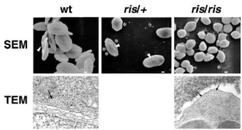
Cell membrane and microtubule marginal band defects of ris erythrocytes. SEM and TEM analysis of peripheral blood cells. Wild-type erythrocytes are elliptical with smooth biconcave membrane surface. A central nuclear bulge (arrow) located within the biconcavity of wildtype erythrocytes corresponds to the nucleus. Heterozygote ris/+ erythrocytes are elliptical, but the membrane surface appears irregular (asterisk) and biconcavity is lost (arrowheads). Homozygote ris/ris red cells are smaller, spherocytic, and show profound surface pitting and spiculated membrane projections (asterisk). TEM analysis at 3000x magnicication, shows that the MB is situated adjacent to the lipid bilayer (arrows). Wild-type or ris/+ (not shown) MB consists of 20-24 microtubules, whereas ris MB consists of only 12-14 microtubule filaments. |