
Fig. 3
- ID
- ZDB-FIG-070116-2
- Publication
- Liao et al., 2000 - Hereditary spherocytosis in zebrafish riesling illustrates evolution of erythroid ß-spectrin structure, and function in red cell morphogenesis and membrane stability
- Other Figures
- All Figure Page
- Back to All Figure Page
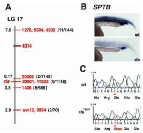
sptb is the defective gene in ris. (A) Genetic map of LG 17, showing a sample number of SSLP markers tested in the meiotic mapping (in red), and their relative distances from ris locus (in black). The number of recombinants/the total number of animals tested are indicated in parenthesis. (B) sptb whole-mount RNA in situ on wild-type and ris embryos at 23 hpf. Identical staining of ICM, myotube and somites is observed with riboprobes generated from BScd, BF1/238 (shown here), or BSPH clones. (C) Sequence of a five codon interval encompassing the C to T transversion mutation at position +3130 that was found in ris. |
| Gene: | |
|---|---|
| Fish: | |
| Anatomical Term: | |
| Stage: | 26+ somites |