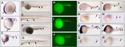
Expression profile of zbp-89 in wild-type zebrafish embryos and the phenotype of zbp-89 morphants. (A-C) Tissue expression of zbp-89 by whole-mount in situ hybridization of 12 hpf (A), 18 hpf (B) and 24 hpf (C) embryos. Lateral views, dorsal upward, anterior to the left are shown. By 12 hpf (A), zbp-89 is expressed in the anterior (arrowhead) and posterior (arrow) lateral plate mesoderm. At 18 hpf (B), it is strongly expressed in the anterior ICM (arrow) and the brain region (arrowhead). At 24 hpf (C), expression in the anterior ICM (arrow) and the wedge region of the anterior ICM (short arrow) is seen together with expression in the brain (arrowhead). No signal was detected with the sense probe (not shown). (D-G) Loss of ZBP-89 results in a bloodless phenotype. DAF staining of 48 hpf whole-mount zebrafish embryos. Blood (arrows) is present in the representative control (mismatch atgMO; D,E) but not in atgMO-injected (F,G) embryos (different embryos are shown in F and G). The bloodless phenotype was present in 78% and 62% of the 100-115 embryos injected at the one- to two-cell stage with either ZBP-89 atgMO or spliceMO, respectively. All views are lateral with anterior left and dorsal top. (H-J). Rescue of zbp-89 morphants by tissue-specific expression of human ZBP-89 under control of the flk1 promoter. Gata1-driven GFP in 22 hpf transgenic (gata1:EGFP) zebrafish embryos that were untreated (H), injected with atgMO only (I) or injected with atgMO plus flk1-ZBP-89 plasmid (J) at the one- to two-cell stage. Single 22 hpf embryos were examined under a fluorescent microscope revealing gata1 expression (red arrows) in the ICM of the untreated (H) and ZBP-89-rescued (J), but not in atgMO only-injected (I), embryos. (K-R) Effect of ZBP-89 knockdown on expression of early hematopoiesis markers. Whole-mount in situ hybridization in wild-type (WT; K,M,O,Q) and ZBP-89-depleted (L,N,P,R) 24 hpf embryos. Embryos were hybridized with digoxigenin-labeled RNA probes for scl (K,L), lmo2 (M,N) and gata2 (O,P). In wild type, scl is expressed in the anterior ICM (K, arrow), posterior ICM (arrowhead in K) and the wedge region of anterior the ICM (K, short arrow). Only minimal expression remains in the wedge region and the posterior ICM in the zbp-89 morphants (L). Lmo2 (M) and gata2 (O) display a similar expression pattern to scl in wild-type embryos, and expression of both is markedly reduced in the ICM of zbp-89 morphants (N and P, respectively). Gata2 is also expressed in the brain and spinal ganglia (short arrows in O,P); its expression at these sites is somewhat reduced by the loss of ZBP-89. (Q,R) Expression of cdx4 in the posterior ICM of 24 hpf wild type (Q) and zbp-89 morphants (R). cdx4 expression is not affected by the loss of ZBP-89.
|
