|
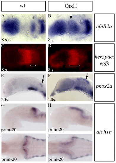
Mesencephalon to rhombomere1 transformation in OtxH embryos. (A-D) Dorsal views (anterior towards the left) of eight-somite embryos after double detection of efnb2a (blue in A,B) expressed in forebrain (f), rhombomeres 1 (r1), 4 (r4) and 7 (r7) and GFP (red in C,D) in wild type; her5pac:egfp (A,C) and OtxH; her5pac:egfp (B,D). In OtxH, the mesencephalon is transformed into an expanded r1 territory (white brackets in C and D indicate the anteroposterior extent of efnb2a). (E-J) Lateral (E-H) and dorsal (I,J) views, anterior towards the left, of wild-type (E,G,I) and OtxH (F,H,J) brains. Locus coeruleus cells expressing phox2a (arrows in E,F) and rhombic lip cells expressing atoh1b (G-J), which is known to arise from the r1 territory, are expanded in OtxH embryos (F,H,J). I and J are dorsal views of embryos in G and H.
|