
FIGURE
Fig. 5
- ID
- ZDB-FIG-051209-1
- Publication
- Prabhudesai et al., 2005 - Targeted effects of retinoic acid signaling upon photoreceptor development in zebrafish
- Other Figures
- All Figure Page
- Back to All Figure Page
Fig. 5
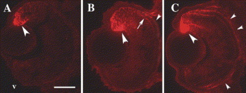
Sectioned zebrafish embryo eyes processed for immunocytochemistry with an anti-RALDH2 (dorsal retinal RA synthesizing enzyme) antibody (red fluorescence). Embryos were fixed at 45 hpf (A), 52 hpf (B), and 80 hpf (C). The antibody labels neuroepithelial cells (large arrowheads) and a few developing neurons, including photoreceptors (arrows), in dorsal retina. At 52 and at 80 hpf, some staining is also evident in ventral retina, throughout the RPE (small arrowheads), and in extraocular locations. v, ventral (in all panels); scale bar = 40 μm. |
Expression Data
| Gene: | |
|---|---|
| Fish: | |
| Anatomical Terms: | |
| Stage Range: | High-pec to Protruding-mouth |
Expression Detail
Antibody Labeling
Phenotype Data
Phenotype Detail
Acknowledgments
This image is the copyrighted work of the attributed author or publisher, and
ZFIN has permission only to display this image to its users.
Additional permissions should be obtained from the applicable author or publisher of the image.
Reprinted from Developmental Biology, 287(1), Prabhudesai, S.N., Cameron, D.A., and Stenkamp, D.L., Targeted effects of retinoic acid signaling upon photoreceptor development in zebrafish, 157-167, Copyright (2005) with permission from Elsevier. Full text @ Dev. Biol.