
FIGURE
Fig. 3
- ID
- ZDB-FIG-070917-134
- Publication
- Prabhudesai et al., 2005 - Targeted effects of retinoic acid signaling upon photoreceptor development in zebrafish
- Other Figures
- All Figure Page
- Back to All Figure Page
Fig. 3
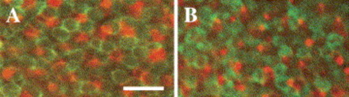
Whole mount double immunocytochemistry using a combination of the zpr-1 (red/green cones) antibody (green fluorescence) and a blue cone opsin antibody (red fluorescence). Regions of whole mounted eye from DMSO-treated control embryos (A) and from RA-treated embryos (B), showing cells stained by each antibody. Embryos were treated at 51 hpf and fixed at 75 hpf. Scale bar = 10 μm. |
Expression Data
Expression Detail
Antibody Labeling
Phenotype Data
Phenotype Detail
Acknowledgments
This image is the copyrighted work of the attributed author or publisher, and
ZFIN has permission only to display this image to its users.
Additional permissions should be obtained from the applicable author or publisher of the image.
Reprinted from Developmental Biology, 287(1), Prabhudesai, S.N., Cameron, D.A., and Stenkamp, D.L., Targeted effects of retinoic acid signaling upon photoreceptor development in zebrafish, 157-167, Copyright (2005) with permission from Elsevier. Full text @ Dev. Biol.