
Fig. 6
- ID
- ZDB-FIG-070917-135
- Publication
- Prabhudesai et al., 2005 - Targeted effects of retinoic acid signaling upon photoreceptor development in zebrafish
- Other Figures
- All Figure Page
- Back to All Figure Page
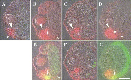
Sectioned eyes derived from embryonic zebrafish, transgenic for a RARE-driven YFP transgene. Embryos were fixed at 45 hpf (A), 52 hpf (B and E), 65 hpf (C and F), and 80 hpf (D and G). (A–D) Immunocytochemistry using an anti-GFP antibody (red fluorescence) shows that neuroepithelial cells (large arrowheads) and a few developing neurons in marginal regions, as well as the RPE (small arrowheads), express the transgene and are therefore likely influenced by RA signaling. Labeling in the dorsal retina decreases after 52 hpf. (E–G) Double immunocytochemistry using the anti-GFP antibody and the zpr-1 (red/green cones) antibody (green fluorescence). Colocalization of the two antibodies (yellow fluorescence) was found in a few ventral photoreceptors at 52 hpf and 80 hpf (arrows). v, ventral (in all panels); scale bar = 40 μm. |
Reprinted from Developmental Biology, 287(1), Prabhudesai, S.N., Cameron, D.A., and Stenkamp, D.L., Targeted effects of retinoic acid signaling upon photoreceptor development in zebrafish, 157-167, Copyright (2005) with permission from Elsevier. Full text @ Dev. Biol.