Effects of Pyripyropene O (PyrO) on the proliferation and migration of prostate cancer cells. (a) Chemical structural formula of Pyripyropene O (PyrO). (b) Effect of PyrO on cell viability. Cell viability was measured by the MTT assay after treatment of prostate cancer cells and normal prostate epithelial cells with different concentrations of PyrO (0–40 μM) for 72 h. (c,d) Effect of PyrO on clone formation in PC-3 cells. PC-3 cells were treated with PyrO (0–10 μM) for 14 days, then stained with crystal violet, and then photographed to observe the effect of PyrO on the ability of PC-3 cells to form clonal colonies. (eg) Effect of PyrO on PC-3 three-dimensional proliferation. After treating PC-3 cell spheres inoculated in U-plates with PyrO (0–40 μM) for 72 h, live and dead cells were stained, and then the size of the cell spheres as well as the number of live and dead cells were observed by fluorescence microscopy. (h,i) Effect of PyrO on PC-3 cell migration. PC-3 cells were inoculated in CIM-plate 16 (5665817001, Agilent) plates, and the cells were treated with PyrO (0–10 μM) for 72 h. The migration of the cells was monitored by real-time cell analysis (RTCA). * p < 0.05, ** p < 0.01 vs. Ctrl.

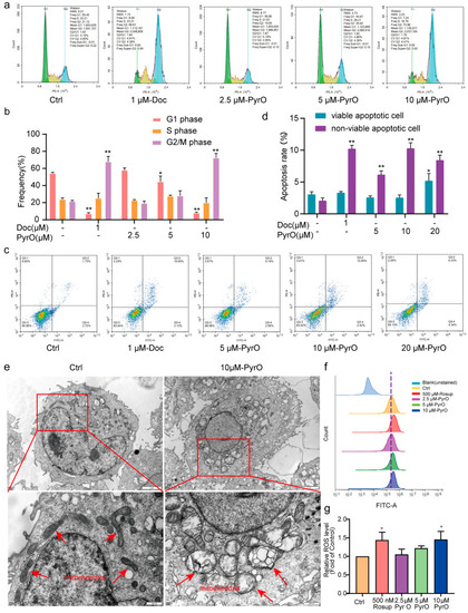
Effects of PyrO on PC-3 cell cycle and apoptosis. (a,b) PyrO blocked PC-3 cells in G2/M phase. Flow cytometry was used to detect changes in the distribution of the PC-3 cell cycle after treatment of PC-3 cells with different concentrations of PyrO for 48 h. (c,d) Effect of PyrO on apoptosis of PC-3 cells. After PyrO (0–20 uM) treatment for 48 h, the cells were stained with Annexin V-FITC/PI, and then the apoptosis was detected by flow cytometry. (e) Effect of PyrO on the internal structure of cells. 10 μM PyrO treated PC-3 cells for 48 h, then the cells were collected to observe the changes in the internal ultrastructure of the cells using transmission electron microscopy to analyze the mode of cell death. The red arrows in the image indicate the location of the mitochondria. (f,g) Effect of PyrO on intracellular ROS levels in PC-3 cells. DCFH-DA fluorescent probe combined with flow cytometry was used to detect the changes in intracellular reactive oxygen species (ROS) levels in PC-3 cells after treatment with different concentrations of PyrO (0–10 μM) for 48 h. * p < 0.05, ** p < 0.01 vs. Ctrl.

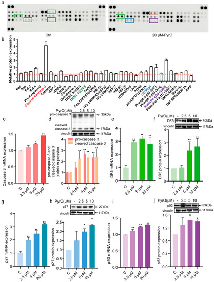
Pyripyropene O regulates the expression of apoptosis-related proteins and their mRNAs in PC-3 cells. (a,b) PC-3 cells were treated with PyrO (20 μM) for 48 h. Cells were collected and apoptosis-associated protein expression changes were detected using protein microarray kit. Red boxes indicate cleaved caspase 3 protein, green boxes indicate TRAIL R2/DR5 protein, blue boxes indicate p27/Kip1 protein, and purple boxes indicate phospho-p53(S392) protein. The red dotted line indicates Baseline (Negative Control). (c) After treating PC-3 cells with different concentrations of PyrO (0–20 μM) for 48 h, total RNA was collected, and the changes in caspase 3 mRNA expression were detected by qPCR after reverse transcription. (d) After treating PC-3 cells with different concentrations of PyrO (0–20 μM) for 48 h, total proteins were collected, and changes in caspase 3 protein expression were detected by western blot. (e) After treating PC-3 cells with different concentrations of PyrO (0–20 μM) for 48 h, total RNA was collected, and the changes in DR5 mRNA expression were detected by qPCR after reverse transcription. (f) After treating PC-3 cells with different concentrations of PyrO (0–20 μM) for 48 h, total proteins were collected, and changes in DR5 protein expression were detected by western blot. (g) After treating PC-3 cells with different concentrations of PyrO (0–20 μM) for 48 h, total RNA was collected, and the changes in p27 mRNA expression were detected by qPCR after reverse transcription. (h) After treating PC-3 cells with different concentrations of PyrO (0–20 μM) for 48 h, total proteins were collected, and changes in p27 protein expression were detected by western blot. (i) After treating PC-3 cells with different concentrations of PyrO (0–20 μM) for 48 h, total RNA was collected, and the changes in p53 mRNA expression were detected by qPCR after reverse transcription. (j) After treating PC-3 cells with different concentrations of PyrO (0–20 μM) for 48 h, total proteins were collected, and changes in p53 protein expression were detected by western blot. * p < 0.05, ** p < 0.01 vs. Ctrl.

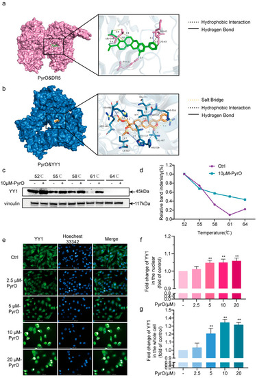
Pyripyropene O targets the YY1/DR5 axis to induce apoptosis. (a) Molecular docking technique was used to reveal the specific binding mode of PyrO to DR5 protein and its intermolecular interactions. (b) Molecular docking technique was used to reveal the specific binding mode of PyrO to YY1 protein and its intermolecular interactions. (c,d) Effect of PyrO on the heat stability of YY1 protein. 10 μM PyrO treated PC-3 for 8 h. Cells were collected, and total protein was collected before analysing the heat stability of the protein using cellular thermal shift assay (CETSA). (eg) Effect of PyrO on nuclear translocation of YY1. PC-3 cells were treated with PyrO for 20 h. Immunofluorescence staining was used to detect the localization of YY1 protein in the cells. ** p < 0.01 vs. Ctrl.

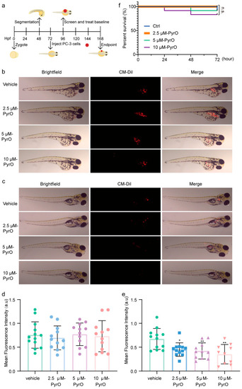
Pyripyropene O inhibits the growth of xenograft prostate cancer PC-3 cells in zebrafish. (a) Procedure for establishing a zebrafish xenograft tumor model. (b) Distribution of prostate cancer PC-3 cells before administration of Pyripyropene O in zebrafish xenografts. (c) Distribution of prostate cancer PC-3 cells in zebrafish after 72 h treatment with different concentrations of Pyripyropene O. (d) Intensity of fluorescence in zebrafish in each group before drug administration. (e) Fluorescence intensity in zebrafish from each group after 72 h of PyrO treatment. (f) Zebrafish survival rate after Pyripyropene O administration. * p < 0.05, ** p < 0.01 vs. vehicle.

Acknowledgments
This image is the copyrighted work of the attributed author or publisher, and ZFIN has permission only to display this image to its users. Additional permissions should be obtained from the applicable author or publisher of the image. Full text @ Mar. Drugs