
- Title
-
Superficial Neurocristic FET::ETS Fusion Tumor: Expanding the Clinicopathological and Molecular Genetic Spectrum of a Recently Described Entity
- Authors
- Dehner, C.A., Warmke, L.M., Umphress, B., Malik, F., Cloutier, J.M., Dermawan, J.K., Fritz, M., Que, S.K.T., Ameline, B., Fritchie, K.J., Kerr, D.A., Linos, K., Baumhoer, D., Billings, S.D., Folpe, A.L.
- Source
- Full text @ Mod Pathol

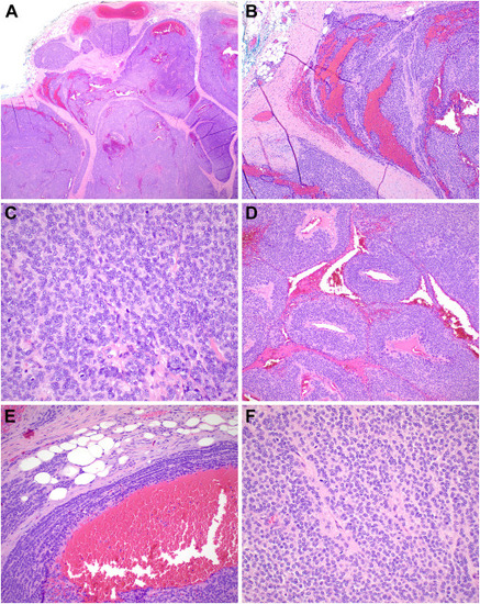
Histopathologic findings of case 1. (A) Low power examination demonstrates a multinodular neoplasm composed of (B) clusters and nests of tumors cells. (C) Foci of the tumor show a distinctive linear arrangement of the tumor cells, whereas other areas (D) demonstrate a nest-like pattern with formation of pseudorosettes. (E) Rare mitotic figures are seen. (F) In some areas, there is a triphasic pattern comprising nests of tumor cells, bland spindled cells and fibroblasts, and scattered adipocytes. (G) S100 protein, (H) SOX10, and (I) CD56 are strongly and diffusely positive. (J) A CD34 stain highlights the outline of the tumor nests giving rise to a vaguely trabecular pattern. (K) CD99 shows weak, membranous staining pattern. |

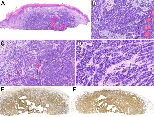
Histopathologic findings of case 2. (A) Low-power histologic examination reveals a dermal-based nodular neoplasm with focal extension into the subcutaneous tissue. (B) Infiltration of adipocytes is noted. (C, D) The tumor cells are epithelioid-to-plasmacytoid with variably prominent eosinophilic cytoplasm. (E) Cytologic atypia is absent. (F) SOX10 is strongly and diffusely positive. (G) CD99 shows membranous staining. |
Histopathologic features of case 3. (A) This tumor presented as deep-dermal to subcutaneous lesion demonstrating a multinodular pattern. (B) Large zones of hemorrhage were present. (C) There was conspicuous mitotic activity. (D) Perivascular growth surrounding vascular spaces was marked in this example. (E) At the periphery, the described triphasic growth pattern was focally noticeable. (F) The tumor cells were bland, round, and monotonous with a vaguely glomoid appearance. |

Histopathologic features of case 4. (A) This tumor also presented as a deep-dermal, somewhat circumscribed lesion with areas of peripheral infiltration (B). (C) At the periphery, the cells often lined up in a single file pattern. (D) Pseudocyst-like spaces with proteinaceous secretions were noted as well as several mitotic figures (E). (F) SOX10 and (G) S100 stains are diffusely positive. |
Histopathologic features of case 5. (A) The superficial shave biopsy of this case showed an infiltrative basaloid dermal neoplasm with multifocal areas of hemorrhage. (B) The growth pattern was predominantly nested, mimicking a melanocytic neoplasm. (C) Peripherally, the cells showed a more spindled growth pattern. (D) Cytologically, the tumor cells were composed of bland, round to ovoid cells with small nucleoli intervened by slightly hyalinized collagen. (E) SOX10 and (F) S100 protein were strongly and diffusely positive. |
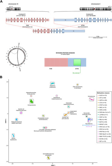
Molecular genetic findings and methylation results. (A) FUS::ETV5 fusion depicted by circos plot, schematic diagram of fusion transcript and predicted chimeric protein with functional domains annotated by National Center for Biotechnology Information RefSeq accession numbers. (B) Uniform Manifold Approximation and Projection (UMAP) plots showing unsupervised clustering of the methylation profiles of all 4 tested tumors in relation to 3 additional superficial neurocristic S100 protein/SOX10-positive tumors, neural tumors, and other mesenchymal neoplasms. Our cases clustered closely with previously reported neurocristic tumors as well as neurofibromas and schwannomas and away from conventional and adamantinoma-like Ewing sarcoma. AFH, angiomatoid fibrous histiocytoma; ALES, adamantinoma-like Ewing sarcoma; ASPS, alveolar soft part sarcoma; CCS, clear cell sarcoma; DFSP, dermatofibrosarcoma protuberan; EMC, extraskeletal myxoid chondrosarcoma; EWS, Ewing sarcoma; ICMT, intracranial mesenchymal tumor; LGFMS, low-grade fibromyxoid sarcoma; MLPS, myxoid liposarcoma; MPNST, malignant peripheral nerve sheath tumor; NFB, neurofibroma; RMS, rhabdomyosarcoma; SCHW, schwannoma; SEF, sclerosing epithelioid fibrosarcoma. |