
Image
|
Figure Caption
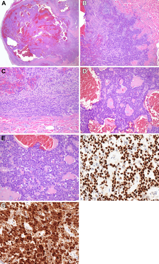
Fig. 4 Histopathologic features of case 4. (A) This tumor also presented as a deep-dermal, somewhat circumscribed lesion with areas of peripheral infiltration (B). (C) At the periphery, the cells often lined up in a single file pattern. (D) Pseudocyst-like spaces with proteinaceous secretions were noted as well as several mitotic figures (E). (F) SOX10 and (G) S100 stains are diffusely positive.
Acknowledgments
This image is the copyrighted work of the attributed author or publisher, and
ZFIN has permission only to display this image to its users.
Additional permissions should be obtained from the applicable author or publisher of the image.
Full text @ Mod Pathol