
FIGURE
Fig. 3
- ID
- ZDB-FIG-250327-68
- Publication
- Dehner et al., 2025 - Superficial Neurocristic FET::ETS Fusion Tumor: Expanding the Clinicopathological and Molecular Genetic Spectrum of a Recently Described Entity
- Other Figures
- All Figure Page
- Back to All Figure Page
Fig. 3
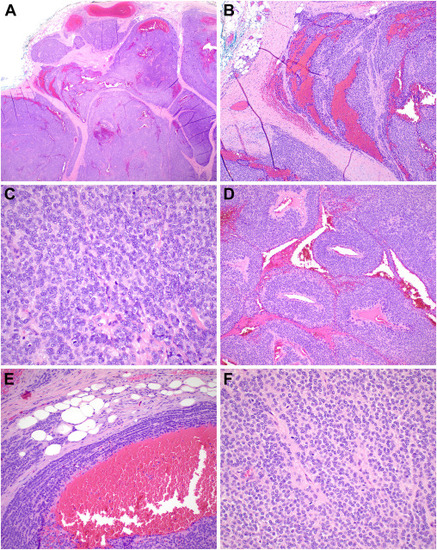
Histopathologic features of case 3. (A) This tumor presented as deep-dermal to subcutaneous lesion demonstrating a multinodular pattern. (B) Large zones of hemorrhage were present. (C) There was conspicuous mitotic activity. (D) Perivascular growth surrounding vascular spaces was marked in this example. (E) At the periphery, the described triphasic growth pattern was focally noticeable. (F) The tumor cells were bland, round, and monotonous with a vaguely glomoid appearance. |
Expression Data
Expression Detail
Antibody Labeling
Phenotype Data
Phenotype Detail
Acknowledgments
This image is the copyrighted work of the attributed author or publisher, and
ZFIN has permission only to display this image to its users.
Additional permissions should be obtained from the applicable author or publisher of the image.
Full text @ Mod Pathol