
Image
|
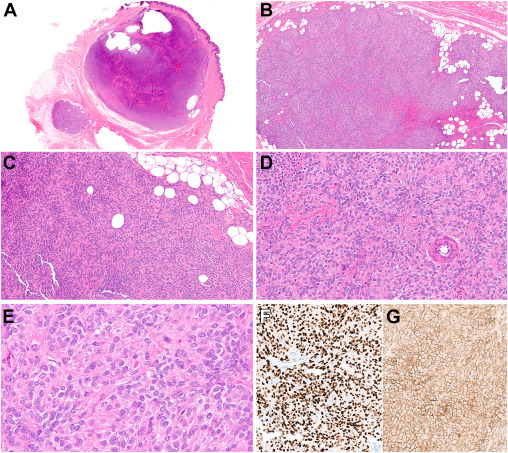
Figure Caption
Fig. 2 Histopathologic findings of case 2. (A) Low-power histologic examination reveals a dermal-based nodular neoplasm with focal extension into the subcutaneous tissue. (B) Infiltration of adipocytes is noted. (C, D) The tumor cells are epithelioid-to-plasmacytoid with variably prominent eosinophilic cytoplasm. (E) Cytologic atypia is absent. (F) SOX10 is strongly and diffusely positive. (G) CD99 shows membranous staining.
Acknowledgments
This image is the copyrighted work of the attributed author or publisher, and
ZFIN has permission only to display this image to its users.
Additional permissions should be obtained from the applicable author or publisher of the image.
Full text @ Mod Pathol