
FIGURE
Fig. 5
- ID
- ZDB-FIG-250327-70
- Publication
- Dehner et al., 2025 - Superficial Neurocristic FET::ETS Fusion Tumor: Expanding the Clinicopathological and Molecular Genetic Spectrum of a Recently Described Entity
- Other Figures
- All Figure Page
- Back to All Figure Page
Fig. 5
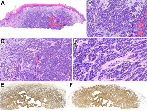
Histopathologic features of case 5. (A) The superficial shave biopsy of this case showed an infiltrative basaloid dermal neoplasm with multifocal areas of hemorrhage. (B) The growth pattern was predominantly nested, mimicking a melanocytic neoplasm. (C) Peripherally, the cells showed a more spindled growth pattern. (D) Cytologically, the tumor cells were composed of bland, round to ovoid cells with small nucleoli intervened by slightly hyalinized collagen. (E) SOX10 and (F) S100 protein were strongly and diffusely positive. |
Expression Data
Expression Detail
Antibody Labeling
Phenotype Data
Phenotype Detail
Acknowledgments
This image is the copyrighted work of the attributed author or publisher, and
ZFIN has permission only to display this image to its users.
Additional permissions should be obtained from the applicable author or publisher of the image.
Full text @ Mod Pathol