
- Title
-
The Xaliproden Nanoscale Zirconium-Porphyrin Metal-Organic Framework (XAL-NPMOF) Promotes Photoreceptor Regeneration Following Oxidative and Inflammatory Insults
- Authors
- Wang, Y., Yuan, B., Liu, W., Cui, J., Zhou, X., Yuan, L., Deng, Z., Li, Y., Yuan, X.
- Source
- Full text @ Int. J. Nanomedicine
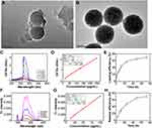
The loading efficiency and release efficiency in NPMOF of XAL. (A) TEM image of NPMOF with an average size of 100 nm. (B) TEM image of XAL-NPMOF. (C) UV‒Vis absorbance and (D) standard curve of XAL in DMF. (E) The loading performance of XAL at 2, 4, 8, 12, 24, 48, and 72 h. (F) Fluorescence and (G) standard curve of XAL in 0.5% hyaluronic acid. (H) The release performance of XAL at 4, 8, 12, 24, 48, and 72 h. Scale bars in (A) and (B), 100 nm. |
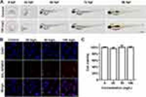
The biological safety of XAL-NPMOF in zebrafish larvae and HUVECs. (A) Phenotypes of larvae after exposure to XAL-NPMOF. (B) Images of HUVECs incubated for 8 h with XAL-NPMOF. Note that the red spots are XAL-NPMOF. (C) Viability of HUVECs cultured with XAL-NPMOF. The dorsal region is up, and the rostral region is left in (A). Scale bar in (A): 500 μm; (B): 20 μm. Abbreviation: hpf, hours post fertilization |
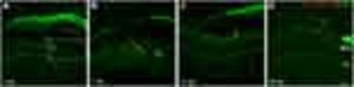
The distribution of XAL-NPMOF in ocular tissue at 3, 7, 14 and 28 dpl. The distribution of XAL-NPMOF (arrowheads) around the lesioned (rectangle) or renewed retina at 3 (A), 7 (B), 14 (C) and 28 dpl (D). Red fluorescence indicates XAL-NPMOF, bright green indicates blood vessels, and dark green indicates background fluorescence. Scale bar: 20 μm. Abbreviations: dpl, days post lesion; CL, choroid layer; RPE, retinal pigment epithelium; PL, photoreceptor layer. |
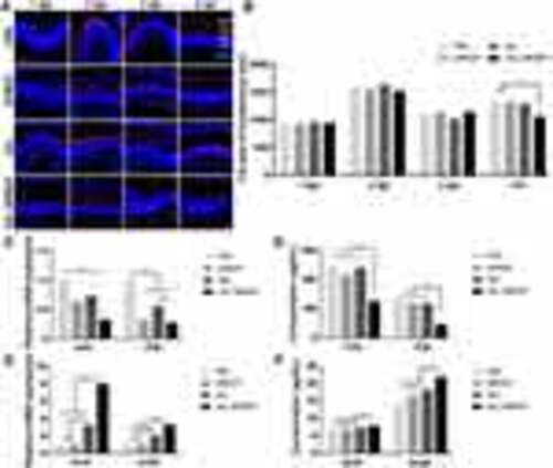
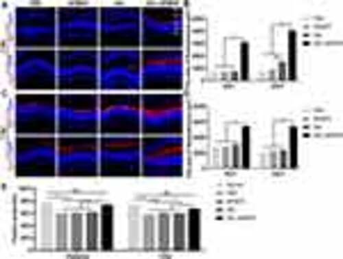
Microglial invasion and anti-inflammatory effects following treatment with XAL-NPMOF. (A) 4C4 staining of sections taken from retinas in the PBS, NPMOF, XAL and XAL-NPMOF groups at 1, 2, 3 and 4 dpl. The 4C4-positive cells are shown in red. (B) Quantification of the area of microglia in the lesioned retina (ANOVA, *p < 0.05). (C) The expression of tnfα and il1β mRNA at 24 hpl in the PBS, NPMOF, XAL and XAL-NPMOF groups (ANOVA, *p < 0.05). (D) Quantification of TNF-α and IL1β expression at 24 hpl via ELISA (ANOVA, *p < 0.05). (E) The expression of sod1 and sod2 mRNA at 24 hpl (ANOVA, *p < 0.05). (F) Quantification of Sod1 and Sod2 expression at 24 hpl via ELISA (ANOVA, *p < 0.05). Scale bar in (A): 50 μm. Abbreviations: dpl, days post lesion; RPE, retinal pigment epithelium; OSL, outer segment layer; ONL, outer nuclear layer; INL, inner nuclear layer; GCL, ganglion cell layer. |
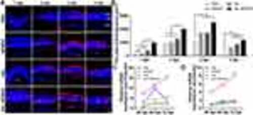
Cell proliferation of phototoxicity-injured retinas following injection of XAL-NPMOF. (A) PCNA staining in sections taken from retinas at 1, 2, 3 and 4 dpl. (B) Statistical analysis of the area of PCNA-positive cells. There was a significantly larger area in the XAL-NPMOF group than in the PBS, NPMOF and XAL groups at 1 and 2 dpl (ANOVA, *p < 0.05). There were more PCNA-positive cells in the retinas of the NPMOF, XAL and XAL NPMOF groups than in those of the PBS group at 3 and 4 dpl (ANOVA, *p < 0.05). (C and D) The expression of ascl1a and sox2 mRNA at 36, 48 and 72 hpl (ANOVA, *p < 0.05). Scale bar in (A): 50 μm. Abbreviations: dpl, days post lesion; hpl, hours post lesion; ONL, outer nuclear layer; INL, inner nuclear layer; GCL, ganglion cell layer. |
Degeneration-regeneration of photoreceptors and changes in visual function following treatment with XAL-NPMOF. (A) Zpr1 and Zpr3 staining in sections taken from retinas of zebrafish in the PBS, NPMOF, XAL and XAL-NPMOF groups at 3 dpl. (B) Quantification of the number of cones and rods in the lesioned retina at 3 dpl. More photoreceptors remained in the XAL-NPMOF group than in the PBS, NPMOF and XAL groups (ANOVA, *p < 0.05). (C) Zpr1 and Zpr3 staining in sections taken from retinas of zebrafish in four groups at 7 dpl. (D) Quantification of the number of cones and rods at 7 dpl. More photoreceptors remained in the XAL-NPMOF group than in the PBS, NPMOF and XAL groups (ANOVA, *p < 0.05). (E) Statistical analysis of the positive correlations of distance and time at 7 dpl. Note that there was a significant decrease in the PBS, NPMOF and XAL groups compared to the normal group in terms of the positive correlation between distance and time (ANOVA, *p < 0.05), while there was no obvious difference between the normal and XAL-NPMOF groups (ANOVA, p > 0.05). Scale bar in (A) and (C): 50 μm. Abbreviations: dpl, days post lesion; RPE, retinal pigment epithelium; OSL, outer segment layer; ONL, outer nuclear layer; INL, inner nuclear layer; GCL, ganglion cell layer. |