
|
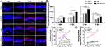
Fig. 5 Cell proliferation of phototoxicity-injured retinas following injection of XAL-NPMOF. (A) PCNA staining in sections taken from retinas at 1, 2, 3 and 4 dpl. (B) Statistical analysis of the area of PCNA-positive cells. There was a significantly larger area in the XAL-NPMOF group than in the PBS, NPMOF and XAL groups at 1 and 2 dpl (ANOVA, *p < 0.05). There were more PCNA-positive cells in the retinas of the NPMOF, XAL and XAL NPMOF groups than in those of the PBS group at 3 and 4 dpl (ANOVA, *p < 0.05). (C and D) The expression of ascl1a and sox2 mRNA at 36, 48 and 72 hpl (ANOVA, *p < 0.05). Scale bar in (A): 50 μm. Abbreviations: dpl, days post lesion; hpl, hours post lesion; ONL, outer nuclear layer; INL, inner nuclear layer; GCL, ganglion cell layer.