
- Title
-
The Increased Burden of Rare Variants in Four Matrix Metalloproteinase-Related Genes in Childhood Glaucoma Suggests a Complex Genetic Inheritance of the Disease
- Authors
- Tevar, A., Aroca-Aguilar, J.D., Bonet-Fernández, J.M., Atienzar-Aroca, R., Campos-Mollo, E., Méndez-Hernández, C., Morales-Fernández, L., Leal Palmer, I., Coca-Prados, M., Martinez-de-la-Casa, J.M., Garcia-Feijoo, J., Escribano, J.
- Source
- Full text @ Int. J. Mol. Sci.
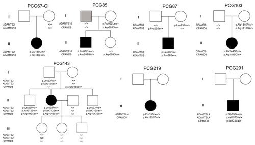
Pedigree and segregation analysis in families of childhood glaucoma patients who carried more than one variant in MMP-related genes. In families PCG219 and PCG291, only proband DNA samples were available for the study. Black and grey symbols indicate childhood glaucoma and primary open-angle glaucoma, respectively. +: wildtype allele. |

Localization of ADAMTSL4 protein in adult human ocular tissues. Fluorescent immunohistochemistry was performed on 3 μm histological sections. The sections were incubated with a primary antibody, rabbit anti-ADAMTSL4 (1:250) (MBS716409, Quimigen, Nanterre, France), followed by incubation with a green-labeled secondary antibody (Cy2-conjugate donkey anti-rabbit) (1:1000). In the resulting images, ADAMTSL4 immunoreactivity, DAPI nuclear staining and tissue autofluorescence are represented by green, blue and red signals, respectively. As a negative control, sections were incubated only with the secondary antibody ( |
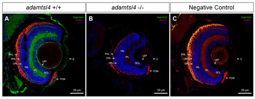
Localization of ADAMTSL4 protein in zebrafish eye larvae (6 dpf) by confocal fluorescence immunohistochemistry. Fluorescent immunohistochemistry was performed on 10 μm histological sections of zebrafish eyes from wild type (+/+) ( |
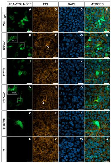
Evaluation of the functional effect of four rare |

Western blot analysis of ER stress associated with the expression in HEK-293T cells of four rare ADAMTSL4 variants identified in childhood glaucoma patients. HEK-293T cells were transfected with various cDNA constructs as explained in the legend of |
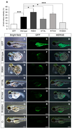
Evaluation of the functional effect of four rare ADAMTSL4 variants identified in childhood glaucoma patients, by heterologous expression in zebrafish. One-cell zebrafish embryos were microinjected with pcDNA3.1-hADAMTSL4-GFP cDNA constructs encoding the four variants (R98W, n = 374; S719L, n = 381; R774W, n = 477; and R1083H, n = 485) and the wild type protein (n = 1013). ( |
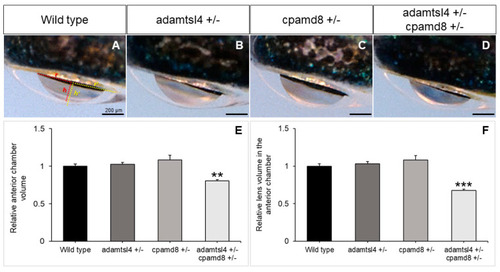
Functional interaction between zebrafish PHENOTYPE:
|