FIGURE

Figure 4

ID
ZDB-FIG-240620-180
Publication
Tevar et al., 2024 - The Increased Burden of Rare Variants in Four Matrix Metalloproteinase-Related Genes in Childhood Glaucoma Suggests a Complex Genetic Inheritance of the Disease
Other Figures
All Figure Page
Back to All Figure Page
Figure 4

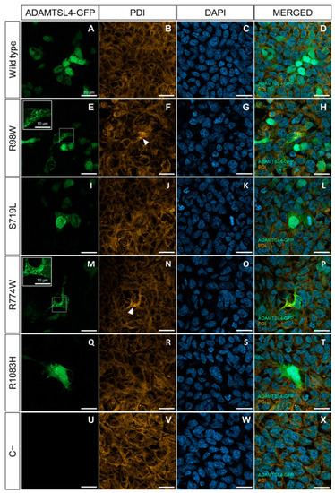
Evaluation of the functional effect of four rare ADAMTSL4 variants identified in childhood glaucoma patients, in HEK-293T cells. Subcellular distribution of the wild type protein (A) and variants R98W (E), S719L (I), R774W (M), and R1083H (Q) in HEK293T Cells (green signals). The cells were transfected with cDNA constructs encoding the different variants, fused to GFP at their C-terminal as a reporter protein and analyzed 48 h after transfection. (U) Non-transfected cells were used as a negative control. (B,F,J,N,R,V) Immunocytochemistry was performed to detect PDI, a marker of ER stress (orange signals). (C,G,K,O,S,W) DAPI nuclear staining (blue signals). (D,H,L,P,T,X) Merged signals. The scale bars in the panels correspond to 20 μm. The white arrowheads indicate increased PDI signal colocalized with the granular expression pattern of the R98W and R774W variants. The inserts in panels (E,M) show granular deposits of recombinant protein, and dotted squares indicate the magnified areas. The images are representative of five fields per variant. Additional representative photographs are shown in Figure S3. R98W: p.Arg98Trp. S719L: p.Ser719Leu. R774W: p.Arg774Trp. R1083H: p.Arg1083His.

Expression Data

Expression Detail
Antibody Labeling
Phenotype Data

Phenotype Detail
Acknowledgments
This image is the copyrighted work of the attributed author or publisher, and ZFIN has permission only to display this image to its users. Additional permissions should be obtained from the applicable author or publisher of the image. Full text @ Int. J. Mol. Sci.