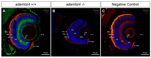
Localization of ADAMTSL4 protein in zebrafish eye larvae (6 dpf) by confocal fluorescence immunohistochemistry. Fluorescent immunohistochemistry was performed on 10 μm histological sections of zebrafish eyes from wild type (+/+) (A) and adamtsl4 KO (−/−) (B) specimens. The sections were incubated with a rabbit anti-ADAMTSL4 primary antibody (MBS716409, Quimigen) at a dilution of 1:250, followed by a Cy2-conjugated donkey anti-rabbit secondary antibody at a dilution of 1:1000. As a negative control (C), a section was incubated only with the secondary antibody. In the resulting images, ADAMTSL4 immunoreactivity is represented by green signals, DAPI nuclear staining by blue signals, and tissue autofluorescence by red signals. Scale bars in the panels indicate 50 μm. The images are representative of the observed results in three zebrafish of each genotype. C: cornea. GCL: ganglion cell layer. INL: inner nuclear layer. IPL: inner plexiform layer. LEP: lens epithelium. ONL: outer nuclear layer. OPL: outer plexiform layer. PHL: photoreceptor layer. POM: periocular mesenchyme.
|
