IMAGE

Figure 2

ID
ZDB-IMAGE-240620-178
Source
Figures for Tevar et al., 2024
Image
Figure Caption

Figure 2

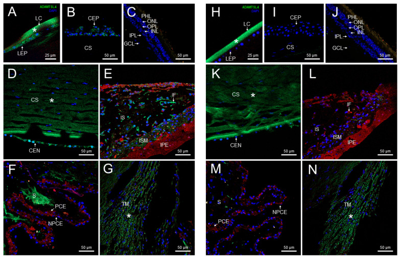
Localization of ADAMTSL4 protein in adult human ocular tissues. Fluorescent immunohistochemistry was performed on 3 μm histological sections. The sections were incubated with a primary antibody, rabbit anti-ADAMTSL4 (1:250) (MBS716409, Quimigen, Nanterre, France), followed by incubation with a green-labeled secondary antibody (Cy2-conjugate donkey anti-rabbit) (1:1000). In the resulting images, ADAMTSL4 immunoreactivity, DAPI nuclear staining and tissue autofluorescence are represented by green, blue and red signals, respectively. As a negative control, sections were incubated only with the secondary antibody (HN). Confocal micrographs were captured to visualize the localization of ADAMTSL4 in different ocular tissues, including the lens (A), corneal epithelium (B), retina (C), corneal endothelium (D), iris (E), ciliary processes (F), and trabecular meshwork (G). The scale bars in panels (BG) correspond to 50 μm, while in panel (A), it corresponds to 25 μm. CEN (corneal endothelium), CEP (corneal epithelium), CS (corneal stroma), GCL (ganglion cell layer), IF (iris fibroblasts), INL (inner nuclear layer), IPL (inner plexiform layer), IPE (iris pigment epithelium), IS (iris stroma), ISM (iris sphincter muscle), LC (lens capsule), LEP (lens epithelium), NPCE (non-pigmented epithelium), ONL (outer nuclear layer), OPL (outer plexiform layer), PCE (pigmented epithelium), PHL (photoreceptor layer), S (stroma), TM (trabecular meshwork). *: non-specific fluorescence.

Acknowledgments
This image is the copyrighted work of the attributed author or publisher, and ZFIN has permission only to display this image to its users. Additional permissions should be obtained from the applicable author or publisher of the image. Full text @ Int. J. Mol. Sci.