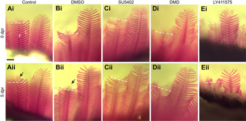
Pharmacological inhibitors restricted regeneration of the gill filaments.In vivo tracking of five individual zebrafish exposed to (A) system water or (B) DMSO as controls, or to (C) SU5402, (D) dorsomorphin (DMD) or (E) LY411575. Gill filaments are shown (Ai–Ei) immediately after the resection procedure and (Aii–Eii) again from the same individual at 5 days post-resection (dpr). In all images, the dashed line indicates the site of resection. Development of a new filament tip (arrows) was observed only in controls (Aii,Bii) at this magnification. F, filament; L, lamella. Scale bar: 100 µm and applies to all panels.
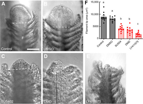
Pharmacological inhibitors reduced the area of gill filament regrowth during regeneration. The distal tips of gill filaments from zebrafish exposed to (A) water or (B) DMSO as controls, or to (C) SU5402, (D) DMD or (E) LY411575 are shown at 5 dpr. A large region of tissue regrowth was observed in controls (A,B) but was reduced in drug treatment groups. In all images, the dashed line indicates the site of resection. F, filament; L, lamella. Scale bar: 50 µm and applies to all panels. (F) Summary data showing means±s.e.m. area of the filament tip (in µm2) at 5 dpr for control and DMSO groups (circles), and for drug treatment groups (squares). Data were analyzed by one-way ANOVA and Tukey's test (F=56.97, P<0.05; N=10 for each group). Significant differences between groups are indicated by different lowercase letters.
Gene expression analysis in regenerating gill filaments using RT-qPCR. (A–E) Relative mRNA expression of fgf8a, bmp2b, bmp6, her6 and jag1b in regenerating gills of zebrafish 10 days after the resection procedure (squares), compared with expression in gills from intact animals (circles). Expression of all genes was observed under control conditions, but relative abundance increased during regeneration for only fgf8a, bmp2b and her6. Data were normalized to the mRNA abundance of the reference gene, ef1a. Data were analyzed using the Mann–Whitney U-test (two-tailed) and means±s.e.m. significantly different from control are indicated by asterisks (U=0, P<0.01; N=8 for each group). n.s., not significant.
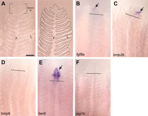
Gene expression in regenerating gill filaments as shown by in situ hybridization. (A) Micrograph of an intact gill filament. In the left panel, the structural filament (F) and numerous respiratory lamellae (L) are shown, as well as the filament tip. In the right panel, the morphology of the gill is outlined for clarity, and approximately corresponds to other panels. The distal tips of previously resected gill filaments at 10 dpr were stained for expression of (B–F) fgf8a, bmp2b, bmp6, her6 and jag1b. Dashed lines indicate the site of resection. Gene expression (arrows) was detected in the regenerating filament tips in B, C and E. Scale bar: 100 µm and applies to all panels.
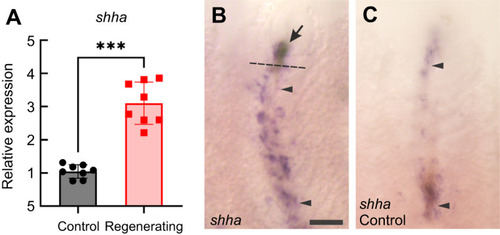
Expression of shha in regenerating gill filaments. (A) Demonstration by RT-qPCR of relative mRNA expression of shha in regenerating gills of zebrafish 10 days after the resection procedure (squares), compared with expression in gills from intact animals (circles). Expression was observed under control conditions, but relative abundance increased during regeneration. Data were normalized to the mRNA abundance of the reference gene, ef1a, and analyzed using the Mann–Whitney U-test (two-tailed). The mean±s.e.m. significantly different from control is indicated by asterisks (U=0, P<0.01; N=8 for each group). (B,C) In situ hybridization demonstrated expression of shha along the length of the filaments (region between arrowheads) in (B) regenerating filaments and in (C) unresected controls, in addition to the increased expression in regenerating tissue (arrow) in B. Scale bar: 100 µm and applies to both panels.
Acknowledgments
This image is the copyrighted work of the attributed author or publisher, and
ZFIN has permission only to display this image to its users.
Additional permissions should be obtained from the applicable author or publisher of the image.
Full text @ J. Exp. Biol.
Your Input Welcome
Thank you for submitting comments. Your input has been emailed to ZFIN curators who may contact you if
additional information is required.
Oops. Something went wrong. Please try again later.