IMAGE

Fig. 1.

ID
ZDB-IMAGE-240304-4
Source
Figures for Cadiz et al., 2023
Image
Figure Caption

Fig. 1.

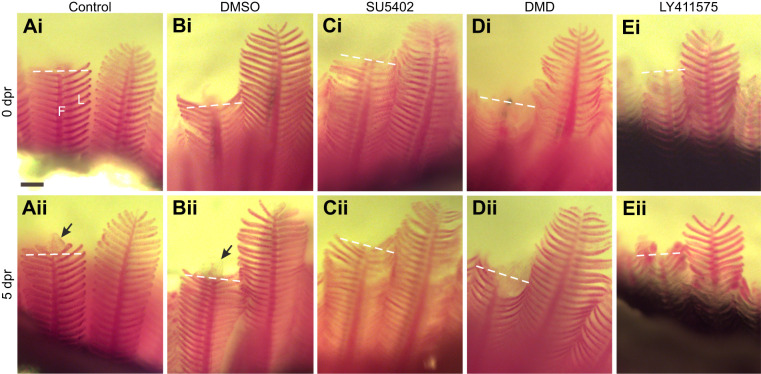
Pharmacological inhibitors restricted regeneration of the gill filaments.In vivo tracking of five individual zebrafish exposed to (A) system water or (B) DMSO as controls, or to (C) SU5402, (D) dorsomorphin (DMD) or (E) LY411575. Gill filaments are shown (Ai–Ei) immediately after the resection procedure and (Aii–Eii) again from the same individual at 5 days post-resection (dpr). In all images, the dashed line indicates the site of resection. Development of a new filament tip (arrows) was observed only in controls (Aii,Bii) at this magnification. F, filament; L, lamella. Scale bar: 100 µm and applies to all panels.

Acknowledgments
This image is the copyrighted work of the attributed author or publisher, and ZFIN has permission only to display this image to its users. Additional permissions should be obtained from the applicable author or publisher of the image. Full text @ J. Exp. Biol.