|
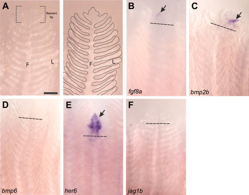
Gene expression in regenerating gill filaments as shown by in situ hybridization. (A) Micrograph of an intact gill filament. In the left panel, the structural filament (F) and numerous respiratory lamellae (L) are shown, as well as the filament tip. In the right panel, the morphology of the gill is outlined for clarity, and approximately corresponds to other panels. The distal tips of previously resected gill filaments at 10 dpr were stained for expression of (B–F) fgf8a, bmp2b, bmp6, her6 and jag1b. Dashed lines indicate the site of resection. Gene expression (arrows) was detected in the regenerating filament tips in B, C and E. Scale bar: 100 µm and applies to all panels.
|