
- Title
-
Tumor-derived exosomal lincRNA ROR promotes angiogenesis in nasopharyngeal carcinoma
- Authors
- Zhang, S., Cai, J., Ji, Y., Zhou, S., Miao, M., Zhu, R., Li, K., Xue, Z., Hu, S.
- Source
- Full text @ Mol. Cell. Probes
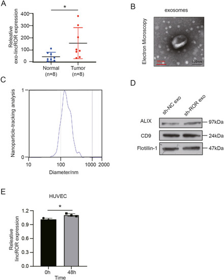
Fig. 1. Serum exosomal linc-ROR expression of NPC patients and healthy donors and the identification of exosomes collected from the cell conditioned medium. Exosomes containing linc-ROR might be picked up by HUVECs. (A) Exo-linc-ROR levels in NPC patients and healthy volunteers detected by qRT-PCR. (B) Transmission electron microscopy image of exosomes (Scale bar: 100 nm). (C) Nanoparticle tracking analysis showed the size distribution of exosomes collected from CNE2 culture medium. (D) Exosomal markers CD9, ALIX, and tubillin-1 were measured by Western blot. (E) qRT-PCR analysis revealed that linc-ROR expression was increased in HUVECs after 48 h of co-culture with exosomes from CNE2 cell conditioned medium. All data show mean ± SD of at least three independent experiments. (*P < 0.05, **P < 0.01, ***P < 0.001, ****P < 0.0001). |
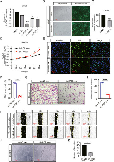
Fig. 2. Elevated exosomal linc-ROR promotes angiogenesis of HUVECs. (A) CNE2 cells were transfected with shROR1, shROR2, shROR3 and a negative control. After 48 h, total RNA from CNE2 cells was isolated and qRT‐PCR for linc-ROR was performed. (B–C) qRT-PCR and fluorescence microscope image (Scale bar: 100 μm) exhibited the transfection efficiency of shROR lentivirus and a negative control lentivirus in CNE2. (D–I) Cell proliferation and migration were detected in HUVECs treated with exosomes from CNE2-NC and CNE2-shROR culture medium. The CCK8 (D) and EDU assay (scale bar: 50 μm) (E, F) were carried out to measure cell proliferation while transwell assays (scale bar: 100 μm) (G, H) and wound healing assay (scale bar: 100 μm) (I) were carried out to measure cell mobility. (J–K) Tube formation assay was used to detect the angiogenesis ability of HUVECs co-cultured with exosomes from CNE2-NC and CNE2-shROR culture medium (scale bar: 100 μm). All data show mean ± SD of at least three independent experiments. (*P < 0.05, **P < 0.01, ***P < 0.001, ****P < 0.0001). |
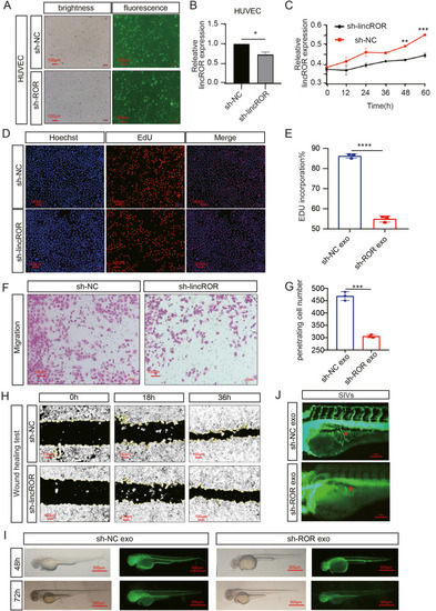
Fig. 3. linc-ROR promotes angiogenesis in vitro and vivo. (A) The fluorescent transfection efficiency of HUVECs was captured by inverted microscope (scale bar: 100 μm) (B) According to qRT-PCR results, linc-ROR level in HUVECs was depressed after transfecting shROR lentivirus compared with transfecting a negative control lentivirus. (C–H) The effect of linc-ROR on proliferation and migration of HUVECs were observed by CCK-8 assay (C), EDU assay (D, E), transwell assays (F, G), wound healing assay (H) (scale bar: 100 μm). (I) Morphology of blood vessels in Tg (flila: EGFP) zebrafish embryos injected with exosomes from CNE2-NC and CNE2-shROR culture medium for 48h, 72h were captured by fluorescence microscope (scale bar: 500 μm). (J) The images displayed were the amplification of SIVs (scale bar: 100 μm). The photographs shown were representative of at least three independent experiments. All data show mean ± SD of at least three independent experiments. (*P < 0.05, **P < 0.01, ***P < 0.001, ****P < 0.0001). |
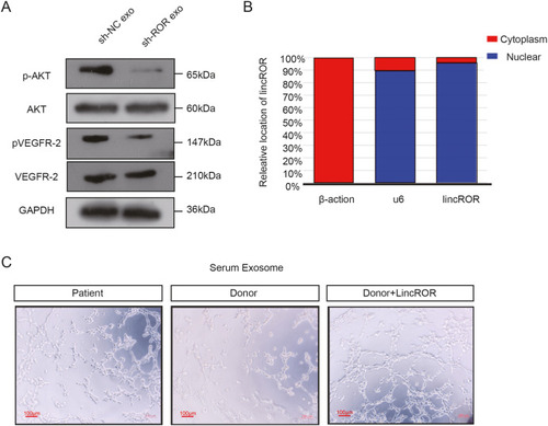
Fig. 4. Exosomal linc-ROR regulates p-AKT/p-VEGFR2 pathway. (A) Western blot analyzed the relative protein level of AKT, p-AKT, VEGFR2, p-VEGFR2 in HUVECs treated with exosomes from CNE2-NC and CNE2-shROR culture medium. (B) The location of linc-ROR was confirmed using qRT-PCR by comparing the expression levels of linc-ROR in the nucleus and cytoplasm. (C) The angiogenesis ability of HUVECs co-cultured with exosomes collected from NPC patients' serum was higher than from healthy donors' serum, while the angiogenesis ability of HUVECs treated with healthy donors' serum exosomes could be enhanced through overexpressing linc-ROR in HUVECs (scale bar: 100 μm). |
Reprinted from Molecular and Cellular Probes, 66, Zhang, S., Cai, J., Ji, Y., Zhou, S., Miao, M., Zhu, R., Li, K., Xue, Z., Hu, S., Tumor-derived exosomal lincRNA ROR promotes angiogenesis in nasopharyngeal carcinoma, 101868, Copyright (2022) with permission from Elsevier. Full text @ Mol. Cell. Probes