
|
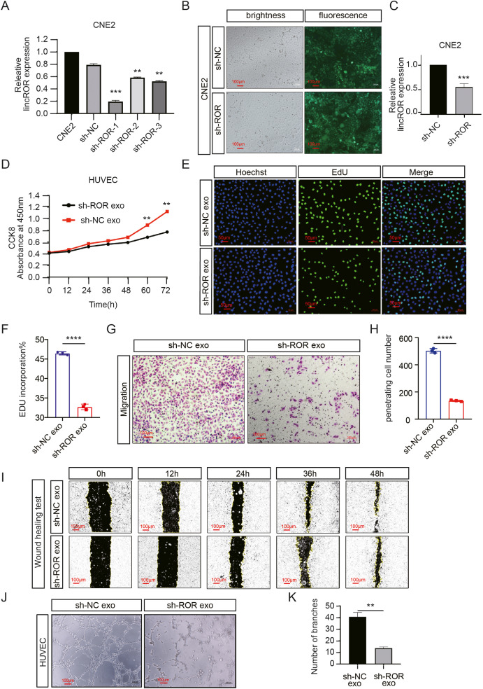
Fig. 2 Fig. 2. Elevated exosomal linc-ROR promotes angiogenesis of HUVECs. (A) CNE2 cells were transfected with shROR1, shROR2, shROR3 and a negative control. After 48 h, total RNA from CNE2 cells was isolated and qRT‐PCR for linc-ROR was performed. (B–C) qRT-PCR and fluorescence microscope image (Scale bar: 100 μm) exhibited the transfection efficiency of shROR lentivirus and a negative control lentivirus in CNE2. (D–I) Cell proliferation and migration were detected in HUVECs treated with exosomes from CNE2-NC and CNE2-shROR culture medium. The CCK8 (D) and EDU assay (scale bar: 50 μm) (E, F) were carried out to measure cell proliferation while transwell assays (scale bar: 100 μm) (G, H) and wound healing assay (scale bar: 100 μm) (I) were carried out to measure cell mobility. (J–K) Tube formation assay was used to detect the angiogenesis ability of HUVECs co-cultured with exosomes from CNE2-NC and CNE2-shROR culture medium (scale bar: 100 μm). All data show mean ± SD of at least three independent experiments. (*P < 0.05, **P < 0.01, ***P < 0.001, ****P < 0.0001).
Reprinted from Molecular and Cellular Probes, 66, Zhang, S., Cai, J., Ji, Y., Zhou, S., Miao, M., Zhu, R., Li, K., Xue, Z., Hu, S., Tumor-derived exosomal lincRNA ROR promotes angiogenesis in nasopharyngeal carcinoma, 101868, Copyright (2022) with permission from Elsevier. Full text @ Mol. Cell. Probes