
- Title
-
Cd248a and Cd248b in zebrafish participate in innate immune responses
- Authors
- Li, X., Guo, R., Yang, S., Zhang, X., Yin, X., Teng, L., Zhang, S., Ji, G., Li, H.
- Source
- Full text @ Front Immunol
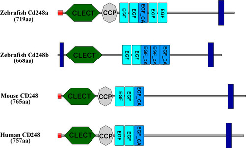
Amino acid functional domains of CD248 protein in human, mouse and zebrafish. Red squares, signal peptide; Blue rectangles, transmembrane region; CLECT, C-type lectin domain; EGF, epidermal growth factor-like domain; EGF_CA, Calcium-binding EGF-like domain; CCP, complement control protein domain. |
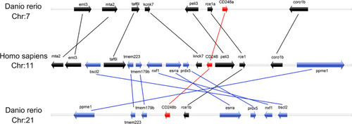
Colinearity analysis between zebrafish |
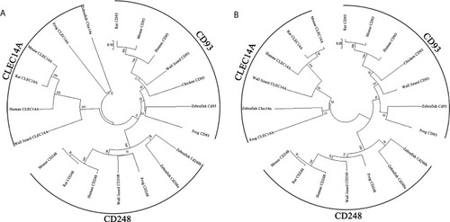
Phylogenetic trees of Cd248a and Cd248b. Phylogenetic trees constructed from the amino acid sequences of homologous genes of various species. CD248, CD93 and CLEC14A are members of the C-type lectin group 14 family. |
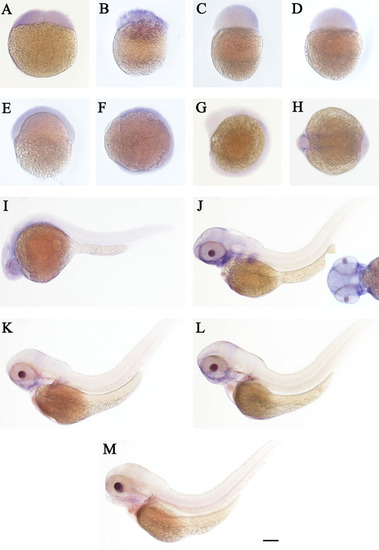
WISH of |
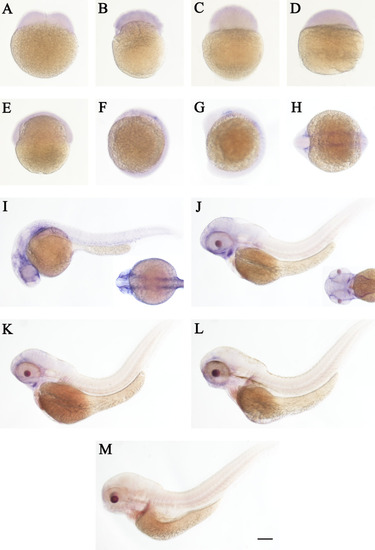
WISH of |
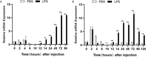
Quantitative analysis of zebrafish |

Subcellular localization of Cd248a, Cd248b and truncated Cd248b in HEK293T cells. The |

Effect of overexpression of zebrafish |
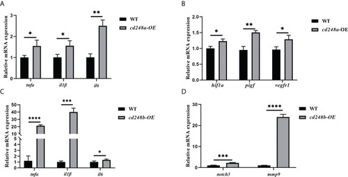
Effect of overexpression of zebrafish |

Effect of |