
- Title
-
Cranberry Pomace Extract Exerts Antiviral Activity against Zika and Dengue Virus at Safe Doses for Adult Zebrafish
- Authors
- Tamkutė, L., Haddad, J.G., Diotel, N., Desprès, P., Venskutonis, P.R., El Kalamouni, C.
- Source
- Full text @ Viruses
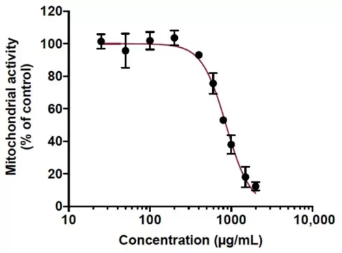
Figure 1. Determination of the maximal non-toxic concentration (MNTC) of cranberry pomace extract on human A549 cell line. A549 cells were grown in the presence of various concentrations of CP extract (25 to 2000 µg/mL) for 48 h. The metabolic activity was assessed by MTT assay. Results presented are means ±SD of three independent experiments and are expressed as relative values to the vehicle control. |
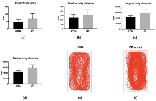
Figure 2. Cranberry pomace extract has no impact on finely tuned locomotor activity one day after injection. (a–c) Distance traveled in the “inactive” (<4 mm/s, (a)), low activity (4–8 mm/s, (b)), and high activity (>8 mm/s, c) states during the 10-min recording period. (d) Total distance traveled by the fish during the recording period. Note that no significant differences were observed between groups. (e,f) Examples of paths traveled by control and CP-injected fish. n = 7 fish/group; data represent means ± SEM. |
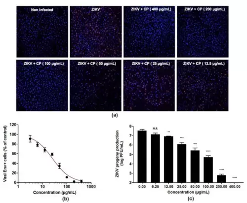
Figure 3. Cranberry pomace extract exhibits dose-dependent antiviral effect against the epidemic strain of ZIKV. A549 cells were challenged with the epidemic strain of ZIKV (PF-25013-18) at an MI of 2 and simultaneously treated with various non-cytotoxic concentrations of CP extract (0–400 µg/mL). (a) Immunofluorescence analysis of viral protein expression in A549-ZIKV-PF13-infected cells. The ZIKV E (red) and nuclei (blue) were visualized by fluorescence microscopy. Images are representative of three independent experiments. (b) Quantification of the number of cells positive for the viral protein E expression in A549-ZIKV-infected cells by immunofluorescence. (c) ZIKV growth was assessed by a plaque formation assay. Results from a representative experiment (n = 3 repeats) are shown. Data are means ± SD of three separate experiments. One-way ANOVA and Dunnett’s test were used for statistical analysis (** p < 0.01; *** p < 0.001; **** p < 0.0001; n.s. = not significant). |
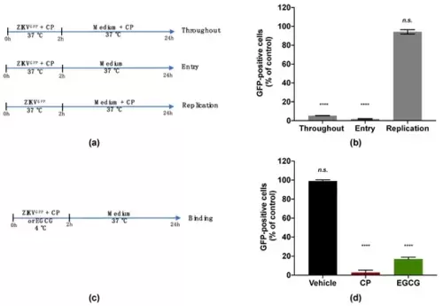
Figure 4. Cranberry pomace extract inhibits the attachment of ZIKV to the surface of A549 cells. (a) Schematic illustration of drug addition time assays applied to describe the antiviral activity of CP extract (100 µg/mL). A549 cells were infected and treated with CP extract or vehicle throughout infection (Throughout), simultaneously with virus entry (Entry), after virus challenge (Replication) with appropriate time-wash steps and incubation periods. (b) Flow cytometric analysis of infected cells under the different experimental conditions shown in (a). (c) Schematic illustration of the binding assay. Pre-chilled ZIKVGFP was mixed with CP extract (100 µg/mL) and left to bind to the A549 cell monolayer at 4 °C for 1 h, then moved to 37 °C. EGCG (100 µM) was used as a positive control. (d) Flow cytometric analysis of cells infected with ZIKVGFP-A549 in the binding assay shown in (c). The results are means ±SD of three independent experiments and are expressed as relative values to vehicle infected cells. One-way ANOVA and Dunnett’s test were used for statistical analysis (**** p < 0.0001; n.s. = not significant). |
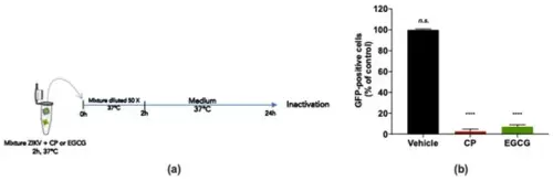
Figure 5. Cranberry pomace extract prevents virus entry by acting directly on ZIKV particles. (a) Schematic illustration of ZIKV inactivation assay performed to characterize the virucidal activity of the CP extract. ZIKVGFP was untreated (vehicle) or treated with the CP extract (100 µg/mL) for 2 h at 37 °C and subsequently diluted 50 times before infection of A549 cells. EGCG (100 µM) was used as a positive control. (b) Flow cytometric analysis under the experimental inactivation conditions shown in (a). The results are means ±SD of three independently performed experiments and are expressed as relative values to untreated infected cells. One-way ANOVA and Dunnett’s test were used for statistical analysis (**** p < 0.0001; n.s. = not significant). |
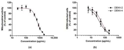
Figure 6. Cranberry pomace extract exhibits antiviral effect against two DENV serotypes. (a) Mitochondrial activity of Huh7.5 cells incubated with different concentrations of CP extract. Cells were grown in the presence of increasing concentrations of CP extract (25 to 2000 µg/mL) for 48 h. The mitochondrial activity of the cells was assessed by MTT assay. (b) Huh7.5 cells were infected for 48 h with DENV-2 or DENV-4 (MI 0.5) in the presence of various non-cytotoxic concentrations of CP extract (6.25–200 µg/mL). Flow cytometric analysis was performed using the anti-flavivirus E mAb 4G2. Data represent means ±SD of three independent experiments. |