
|
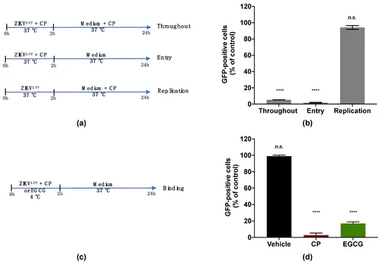
Fig. 4 Figure 4. Cranberry pomace extract inhibits the attachment of ZIKV to the surface of A549 cells. (a) Schematic illustration of drug addition time assays applied to describe the antiviral activity of CP extract (100 µg/mL). A549 cells were infected and treated with CP extract or vehicle throughout infection (Throughout), simultaneously with virus entry (Entry), after virus challenge (Replication) with appropriate time-wash steps and incubation periods. (b) Flow cytometric analysis of infected cells under the different experimental conditions shown in (a). (c) Schematic illustration of the binding assay. Pre-chilled ZIKVGFP was mixed with CP extract (100 µg/mL) and left to bind to the A549 cell monolayer at 4 °C for 1 h, then moved to 37 °C. EGCG (100 µM) was used as a positive control. (d) Flow cytometric analysis of cells infected with ZIKVGFP-A549 in the binding assay shown in (c). The results are means ±SD of three independent experiments and are expressed as relative values to vehicle infected cells. One-way ANOVA and Dunnett’s test were used for statistical analysis (**** p < 0.0001; n.s. = not significant).