FIGURE SUMMARY
Title

MicroRNA-22 coordinates vascular and motor neuronal pathfinding via sema4 during zebrafish development

Authors
Sheng, J., Gong, J., Shi, Y., Wang, X., Liu, D.
Source
Full text @ Open Biol.

Deficiency of miR-22a caused aberrant vascular networks. (a) Confocal imaging analysis of control MO, miR-22a-MO and miR-22a-sponge-injected Tg(kdrl:EGFP) embryos at 30 hpf and 48 hpf. The arrowheads indicate the aberrant angiogenic sprouts. A: dorsal aorta; V: posterior cardinal vein. (b) Statistics of horizontal sprouts ratio in 30 hpf embryos injected with control MO (n = 7), miR-22-MO (n = 7) and miR-22-sponge (n = 7). One-way ANOVA, **** p < 0.0001. (c) Statistics of embryos with vessel across segments ratio in each group: control MO (n = 6), miR-22a-MO (n = 8) and miR-22-sponge (n = 8). One-way ANOVA, ** p < 0.01; **** p < 0.0001. (d) Confocal imaging analysis of control and miR-22a-KO Tg(fli1a:EGFP-CAAX) embryos at 48 hpf. (e) The statistics of embryos with vessels across segments ratio in each group: control (n = 7) and miR-22-KO (n = 7). t-test, ****, p < 0.0001.

miR-22a regulates ISV tip cell behaviour. (a,b) Still images from in vivo time-lapse imaging analysis of Tg(kdrl:EGFP) embryos with HD detection setting. Time stages (hpf) are noted at the left side. The yellow arrows indicate the upward tip cells in the control groups. The blue arrows indicate the lateral tip cells. The red arrows indicate the downward tip cells. The rectangle box in dash line indicates the multidirectional filopodial extensions in the miR-22a deficiency embryos. (c,d) Diagrams of ISV morphology in control and miR-22a MO-injected embryos.

Deficiency of mir-22a caused aberrant axonal projection of PMNs. (a) Confocal imaging analysis of primary motor neurons in the control embryos, miR-22a morphants and Tg(mnx1:GFP::ubi:miR-22a-sponge) embryos at 48 and 72 hpf. Arrowheads indicate the CaPs across different segments. (b,c) Schematic diagram for primary motor neurons in the control embryos and miR-22a morphants. (d) Statistical analysis of the ratio of CaPs across different segments in the control (n = 8), miR-22a morphants (n = 8) and Tg(mnx1:GFP::ubi:miR-22a-sponge) embryos (n = 8) at 48 and 72 hpf; One-way ANOVA, ****p < 0.0001. (e) Statistical analysis of the ratio of aberrant axonal projection of CaPs in the control (n = 8), miR-22a morphants (n = 8) and Tg(mnx1:GFP::ubi:miR-22a-sponge) embryos (n = 8) at 48 and 72 hpf; one-way ANOVA, ****p < 0.0001.

miR-22a regulates primary motor neuron and ISV pathfinding. (a) Confocal imaging analysis of fli1a:miR-22a-sponge-injected Tg(mnx1:EGFP::kdrl:ras-mCherry) embryos at 72 hpf. (b) Percentage of embryos with impaired ISV and PMN pathfinding phenotype in fli1a:miR-22a-sponge-injected embryos at 48 hpf and 72 hpf, respectively (n = 108; n = 120). (c) Confocal imaging analysis of huc:miR-22a-sponge-injected Tg(mnx1:EGFP::kdrl:ras-mCherry) embryos at 72 hpf. (d) Percentage of embryos with indicated phenotypes in huc:miR-22a-sponge-injected embryos at 48 hpf and 72 hpf, respectively (n = 112; n = 105). (d) Quantitative PCR analysis of miR-22a in control neuron cells and neuron cells sorted from embryo injected with ECs expressing miR-22a-sponge at 24 hpf (n = 3), 48 hpf (n = 3), and 72 hpf (n = 3). t-test; **p < 0.001.

miR-22a directly targets sema4c. (a) Venn diagram of predicted target genes of miR-22a and transcriptomic upregulated genes. (b) Sema4c 3′-UTR target sites of miR-22a. (c) Overexpression of miR-22 (miR-22-pre) reduced sema4c-3′-UTR luciferase activity in HeLa cells (n = 9). Data are expressed as mean ± s.e. t-test; **p < 0.01. (d) EGFP sensors were co-injected with mCherry control as indicated. miR-22a-precursor injection reduced the EGFP levels in EGFP-sema4c-3′-UTR sensor (second column), whereas mCherry levels were unchanged. In the mutated sensor, no reduction in GFP was noted (experiments were repeated three times; for each group, around 10 embryos were analysed).

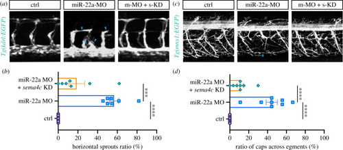
Reducing sema4c partially restored the defects of ISVs and PMNs in miR-22a-deficient embryos. (a) Confocal imaging analysis of blood vessels in control, miR-22a-MO, and miR-22a-MO + sema4c knockdown embryos at 30 hpf. (b) Statistics of horizontal sprouts ratio in control (n = 7), miR-22a-MO (n = 7) and miR-22a-MO + sema4c knockdown embryos (n = 7) at 30 hpf. One-way ANOVA; ***p < 0.001; ****p < 0.0001. (c) Confocal imaging analysis of control, miR-22a-MO and miR-22a-MO + sema4c knockdown Tg(mnx1:EGFP) embryos at 72 hpf. (d) The statistical analysis of the ratio of aberrant axonal projection of CaPs in the control (n = 8), miR-22a morphants (n = 8) and miR-22a-MO + sema4c knockdown embryos (n = 8) at 72 hpf; one-way ANOVA, ****p < 0.0001.

Endothelial miR-c22a regulates PMNs axonal navigation. (a) A possible working model for how blood vessels regulate primary motor neuronal pathfinding in zebrafish. (b) Q-PCR analysis of the miR-22 expression in the isolated exosome from HUVECs (n = 6). (c) Imaging analysis of Tg(mnx1:EGFP::fli1a:CD61-mCherry); arrowhead indicates the vesicle from endothelial cells. (d) Confocal imaging analysis of control, rab11bb MO injected and GW4869 treated Tg(mnx1:EGFP) embryos. (e) Percentage of embryos with indicated phenotypes in control (n = 30), rab11bb MO injected (n = 55) and GW4869 treated Tg(mnx1:EGFP) embryos (n = 38). (f) Percentage of embryos with indicated phenotypes in control (n = 18), miR-22 MO injected (n = 25), miR-22 MO co-injected with exosome isolated from HUVECs (n = 28), miR-22 MO co-injected with exosome isolated from HUVECs transfected with miR-22 duplex (n = 32) and miR-22 MO co-injected with exosome isolated from HUVECs transfected with miR-22 MO Tg(mnx1:EGFP) embryos (n = 23). Fisher's exact test, ****p < 0.0001; ***p < 0.001; ns, no significance.

Acknowledgments
This image is the copyrighted work of the attributed author or publisher, and ZFIN has permission only to display this image to its users. Additional permissions should be obtained from the applicable author or publisher of the image. Full text @ Open Biol.