
- Title
-
Elovl2 Is Required for Robust Visual Function in Zebrafish
- Authors
- Dasyani, M., Gao, F., Xu, Q., Van Fossan, D., Zhang, E., F M Pinto, A., Saghatelian, A., Skowronska-Krawczyk, D., Chao, D.L.
- Source
- Full text @ Cells
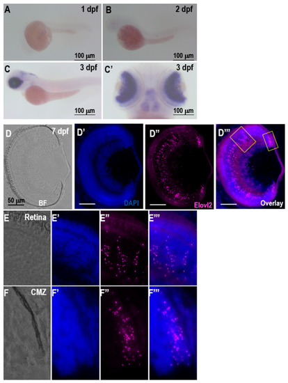
A–C, expression of EXPRESSION / LABELING:
|
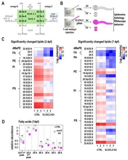
Generation and analysis of |
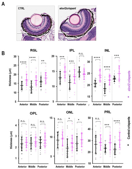
( PHENOTYPE:
|
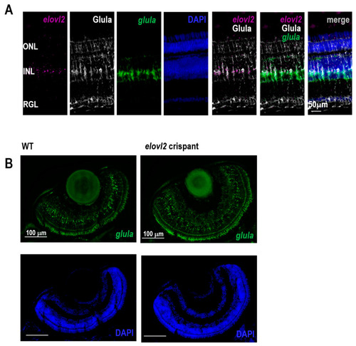
( EXPRESSION / LABELING:
|

Zebrafish PHENOTYPE:
|