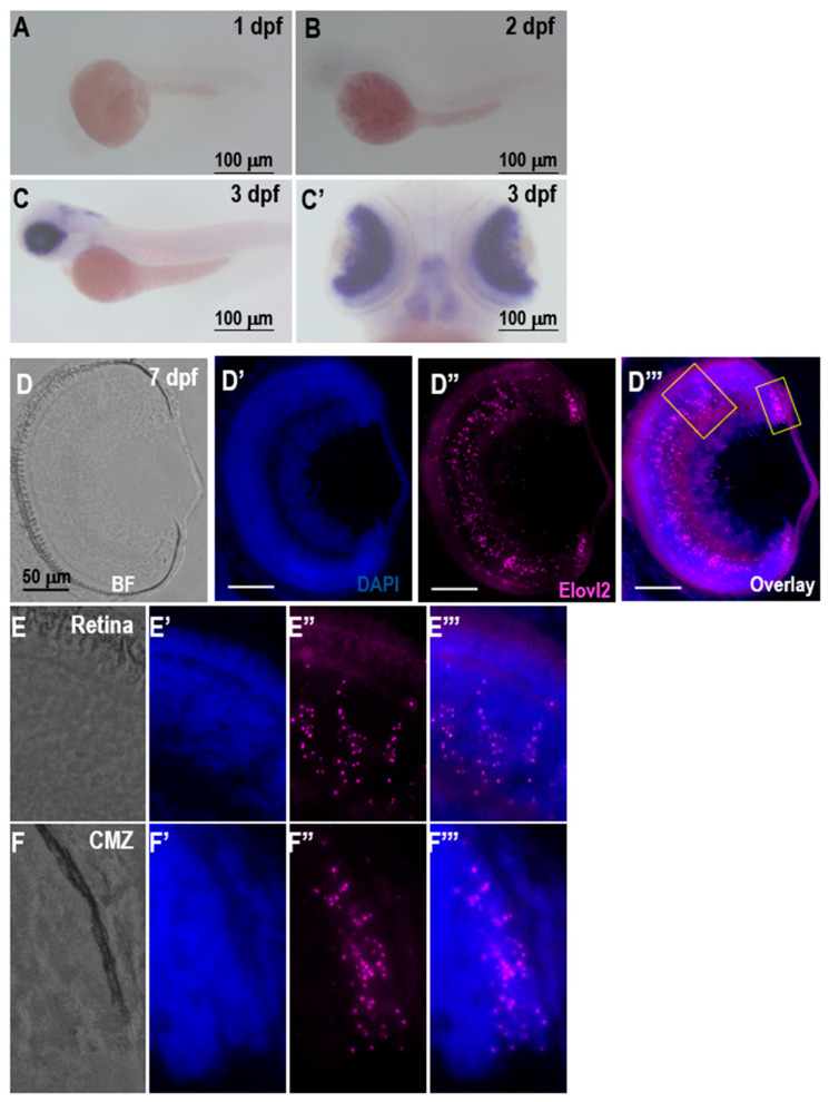
A–C, expression of elovl2 in zebrafish larvae. Lateral view of elovl2 staining in (A) 1 dpf and (B) 2 dpf zebrafish retinas. Lateral (C) and dorsal (C’) view of elovl2 staining in 3 dpf zebrafish retina, respectively. D–F. RNAscope in situ hybridization with elovl2 probe in 7 dpf zebrafish retina showing brightfield (D–F), 4′,6-diamidino-2-phenylindole (DAPI staining (D’–F’), elovl2 (D’’–F’’) and overlay (D’’’–F’’’) images. Scale bars = 50 µm. Magnified view (40×) of boxed regions in Figure 1D’’’. The magnified views of Retina (Panel E) and ciliary marginal zone (CMZ) (Panel F) showing elovl2 staining in 7 dpf zebrafish retina.
This image is the copyrighted work of the attributed author or publisher, and
ZFIN has permission only to display this image to its users.
Additional permissions should be obtained from the applicable author or publisher of the image.
Full text @ Cells
Your Input Welcome
Thank you for submitting comments. Your input has been emailed to ZFIN curators who may contact you if
additional information is required.
Oops. Something went wrong. Please try again later.