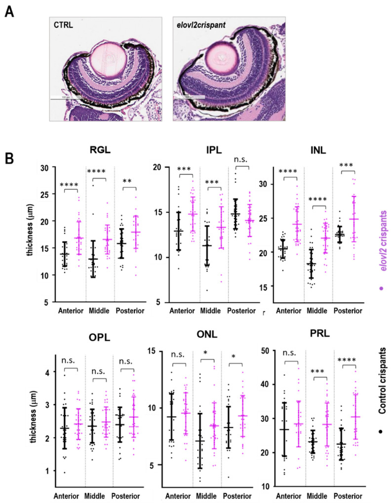
(A) H&E stained retinal sections of elovl2 and control crispants. (B) The thickness of the individual retinal layers of elovl2 and control crispants. Plots showing comparison of lengths of different layers in retina between control and elovl2 crispants. The measurements were taken at three different regions in the retina termed as anterior, posterior, and middle. RGL, Retinal Ganglion Cell Layer; IPL, Inner Plexiform Layer; INL, Inner Nuclear Layer; OPL, Outer Plexiform Layer; ONL, Outer Nuclear Layer; PRL, Photoreceptor Layer. (* p < 0.05; ** p < 0.01; *** p < 0.001; **** p < 0.0001, ns—not significant). Each dot is a single cryosection.
This image is the copyrighted work of the attributed author or publisher, and
ZFIN has permission only to display this image to its users.
Additional permissions should be obtained from the applicable author or publisher of the image.
Full text @ Cells
Your Input Welcome
Thank you for submitting comments. Your input has been emailed to ZFIN curators who may contact you if
additional information is required.
Oops. Something went wrong. Please try again later.