
- Title
-
Anti-Müllerian hormone (Amh/amh) plays dual roles in maintaining gonadal homeostasis and gametogenesis in zebrafish
- Authors
- Zhang, Z., Zhu, B., Chen, W., Ge, W.
- Source
- Full text @ Mol. Cell. Endocrinol.

ZFIN is incorporating published figure images and captions as part of an ongoing project. Figures from some publications have not yet been curated, or are not available for display because of copyright restrictions. EXPRESSION / LABELING:
|
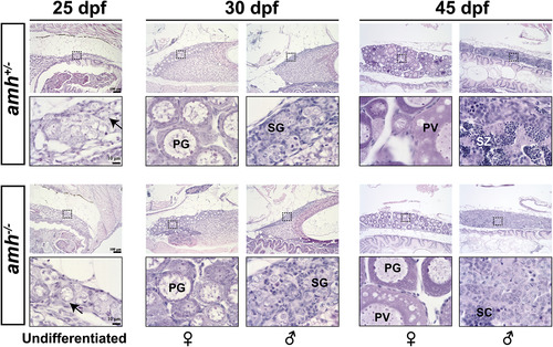
Histological analysis of early gonadal development in the amh mutant. The gonads at 25 dpf were mostly undifferentiated in both amh control (amh+/−) and mutant (amh−/−), whereas well-developed perinucleolar PG follicles and clustered pre-meiotic spermatogonia were found at 30 dpf in the ovary and testis respectively. At 45 dpf, puberty onset had started in females as evidenced by the PV follicles containing cortical alveoli, and spermatogenesis in the testes had entered meiotic phase with large number of spermatocytes and even mature spermatozoa. The arrows indicate the oocyte-like germ cells. PG, primary growth follicles; PV, pre-vitellogenetic follicles; SG, spermatogonia in cysts; SC, spermatocytes; SZ, spermatozoa. PHENOTYPE:
|

Hypertrophic gonads of amh mutants at 4 mpf. (A) The mutant male individuals (amh−/−) showed enlarged abdomen and huge testes (arrows) when compared with the controls (amh+/+ and amh+/−). By comparison, no obvious difference was observed among different genotypes in females. (B) Dissected gonads and quantification of gonadal weight and GSI. *** P < 0.001. (C) Histological examination of testes from different genotypes (amh+/+, amh +/− and amh−/−). The mutant testis (amh−/−) contained much higher number of pre-meiotic spermatogonia (asterisks) but much less meiotic spermatocytes (SC) and post-meiotic mature spermatozoa (SZ). PHENOTYPE:
|
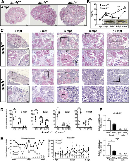
Histologic examination of ovaries from different genotypes (amh+/+, amh +/− and amh−/−) and fecundity analysis. (A) Histology of the ovaries at 4 mpf. Representative sections of the whole ovaries from different genotypes are shown. The mutant ovary (amh−/−) contained much more PG follicles as compared with the controls (amh+/+ and amh+/−). (B) GSI in the control (amh+/−) and mutant (amh−/−) females at different time points (3–12 mpf). The mutants demonstrated higher GSI values after 3 mpf compared with the control (n = 8). The mutant females also showed enlarged abdomen at 12 mpf. (C) Receding folliculogenesis in female amh mutants. Histological analysis of the control (amh+/−) and mutant (amh−/−) ovaries from 2 to 12 mpf. The boxed areas were shown at higher magnification below. PG (stage I), primary growth follicles; PV (stage II), previtellogenic follicles; FG (stage III), full-grown follicles. (D) Analysis of follicle composition in the control (amh+/−) and mutant (amh−/−) ovaries. Two distant sections from each individual were chosen for follicle counting, and the numbers of PG, PV and EV-FG (stage III) follicles in each section were counted. The average of the two sections for each follicle stage was calculated to represent the individual (n = 5). (E) Mating performance and fecundity of the control (amh+/−) and mutant (amh−/−) females. Mating performance is defined as the percentage of successful spawning in each test (n = 8; 15 tests), and fecundity is defined as the number of eggs ovulated and released (n = 8). (F) Expression of vitellogenin genes (vtg1, 2, 4–7, and vtg3) in the liver of control (amh+/−) and mutant (amh−/−) (>4 mpf). * P < 0.05; ** P < 0.01. N.D., not detectable. PHENOTYPE:
|
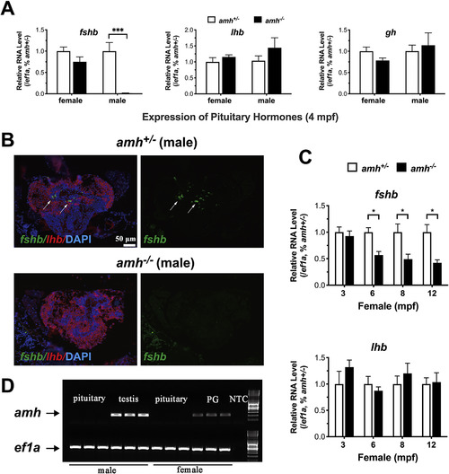
Expression of pituitary hormones (fshb, lhb and gh) in the control (amh+/−) and mutant (amh−/−). (A) Real-time qPCR quantification of fshb, lhb and gh expression (n = 6). (B) Detection of fshb and lhb expression in male pituitaries by fluorescent in situ hybridization. The fshb-expressing cells (green) are indicated by arrows. (C) Expression of fshb and lhb in female pituitaries at 3, 6, 8 and 12 mpf (n = 6). (D) Detection of amh expression in the pituitary and gonads (ovary and testis). * P < 0.05; *** P < 0.001. PG, primary growth follicle; NTC, no template control. (For interpretation of the references to color in this figure legend, the reader is referred to the Web version of this article.) |

Evidence for the involvement of gonadal inhibin in the reduced expression of fshb in the pituitaries of amh mutants. (A) Expression of inha in the ovary and testis of control (amh+/−) and amh mutant (amh−/−) at 4 mpf. The copy numbers of target transcripts (amh and ef1a) were determined by q-PCR with standard curves. The abundance of inha was expressed either as the ratio to that of the housekeeping gene ef1a (upper) or the total amount per gonad (lower). *** P < 0.001 (n = 6). (B) Restored expression of fshb in the double mutant amh−/−;inha−/− at 6 mpf in both females (upper) and males (lower). Different letters indicate statistical significance. (C) Histological examination of the ovaries (upper) and testes (lower) of the amh−/−;inha−/− double mutant at 6 mpf. |
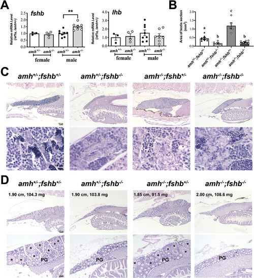
Expression of pituitary gonadotropins (fshb and lhb) and gonadal development in juveniles. (A) Expression of fshb and lhb in the control (amh+/−) and mutant (amh−/−) fish at 45 dpf. (B) Quantification of testis size at 50 dpf. Each fish was sectioned serially and longitudinally and the section that showed the largest testis area was chosen for quantification by the ImageJ software (mm2). Different letters indicate statistical significance (P < 0.05; n = 5–8). The representative section of each genotype is shown in C. (C) Testes of different genotypes at 50 dpf. Compared to the control (amh+/−;fshb+/−), the fshb deficiency (amh+/−;fshb−/−) led to hypotrophy of testis and delayed spermatogenesis, whereas amh deficiency (amh−/−;fshb+/−) led to hypergrowth of testis and dysfunctional spermatogenesis. However, double mutation of fshb and amh (amh−/−;fshb−/−) showed antagonistic effects on testis growth with dysfunctional spermatogenesis. (D) Ovaries of different genotypes at 50 dpf. Mutation of fshb delayed PG-PV transition or puberty onset, whereas amh mutation had no effect. The standard body length (cm) and body weight (mg) are indicated for each fish. PG, primary growth follicles; Asterisk, PV follicles; ** P < 0.01. EXPRESSION / LABELING:
PHENOTYPE:
|
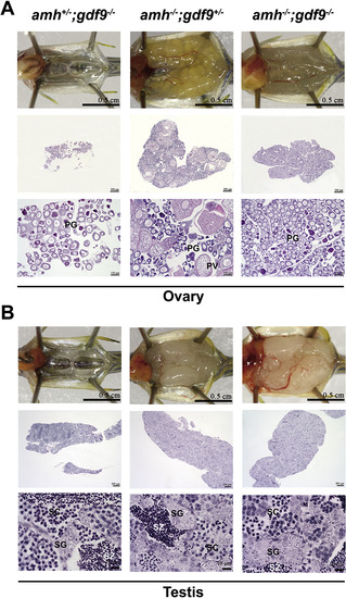
Effects of amh and gdf9 double mutations (amh−/−;gdf9−/−) on gonadal development at 3 mpf (90 dpf). (A) Ovaries of different genotypes. While amh mutation had no effect on PG-PV transition despite increased PG follicles, the mutation of gdf9 completely blocked the transition with all follicles arrested at PG stage. Double mutation of the two genes (amh−/−;gdf9−/−) resulted in enlarged ovaries full of PG follicles only. (B) Testes of different genotypes. The mutation of gdf9 (amh−/−;gdf9−/−) had no effect on testis hypergrowth and dysfunctional spermatogenesis induced by amh single mutation (amh−/−;gdf9+/−). PG, primary growth follicles; PV, pre-vitellogenetic follicles; SG, spermatogonia in cysts; SC, spermatocytes; SZ, spermatozoa. PHENOTYPE:
|

Histological analysis of double and triple knockouts of amh, gdf9 and fshb genes at 3.5 mpf. While double mutation of amh and gdf9 (amh−/−;gdf9−/−;fshb+/−) caused ovarian hypergrowth with PG follicles only, the loss of fshb in the triple knockout (amh−/−;gdf9−/−;fshb−/−) reduced the ovarian size. The ovaries are outlined by dotted lines. |

Amh deficiency attenuated the female-to-male sex reversal in the fshr mutant. (A) Sex ratios in the amh and fshr double mutant (amh−/−;fshr−/−) at 90 and 120 dpf respectively (two different batches of fish). The number of fish analyzed is shown on top of each column. * P < 0.05; ** P < 0.01 compared to the expected sex ratio (1:1) by Chi-square test. (B) Evidence for sex reversal in the double mutant (amh−/−;fshr−/−). Two representative fish had ovotestis structure with ovarian (O) and testis (T) tissues. Note the hypotrophic ovarian tissue and hypertrophic testicular tissue in both individuals. PHENOTYPE:
|
Reprinted from Molecular and Cellular Endocrinology, 517, Zhang, Z., Zhu, B., Chen, W., Ge, W., Anti-Müllerian hormone (Amh/amh) plays dual roles in maintaining gonadal homeostasis and gametogenesis in zebrafish, 110963, Copyright (2020) with permission from Elsevier. Full text @ Mol. Cell. Endocrinol.