
- Title
-
Optic nerve regeneration in larval zebrafish exhibits spontaneous capacity for retinotopic but not tectum specific axon targeting
- Authors
- Harvey, B.M., Baxter, M., Granato, M.
- Source
- Full text @ PLoS One
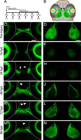
Regeneration of RGC axons in larval zebrafish. (A) Timeline of optic nerve regeneration. (B) Diagram of a Tg(isl2b:GFP) larva expressing GFP in RGCs and their axons. At 5dpf, optic nerves are transected with a sharpened tungsten needle proximal to the chiasm (black arrowheads). (C-D) Before injury, RGC axons cross the midline at the optic chiasm and innervate the contralateral tecta. Dashed lines indicate the outline of the eyes; scale bars = 50 μm. (E-F) Axonal growth from transected nerve at 16hpt is undetectable, while the portion of the nerve distal to the injury degenerates. (G-H) At 24 hpt, regrowing RGC axons begin to emerge from the proximal portion of the injured nerve (white arrowheads), while further degeneration continues. (I-J) By 48 hpt, regenerating axons project into the chiasm and re-innervate the tecta, though about 50% of transected nerves exhibit some misguided axonal growth (n = 81/160 nerves from 80 larvae, white arrows). (K-L) At 72 hpt, there is additional axonal growth to the tecta. (M-N) By 96 hpt, the axons within the chiasm fasciculate and robustly innervate the tecta (n = 160/160 nerves from 80 larvae). Representative images of chiasms and tecta for each timepoint are of the same fixed larva, though across timepoints are different larvae. |
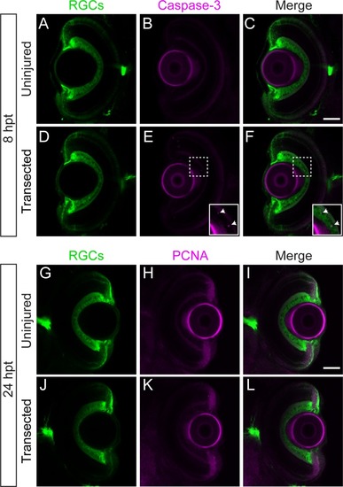
Optic nerve regeneration occurs in absence of significant RGC apoptosis or proliferation. Optic nerve regeneration occurs in absence of significant RGC apoptosis or proliferation. (A-F) Transverse optical sections of retinas from Tg(isl2b:GFP) uninjured larvae (A-C; n = 8 retinas) or larvae with transected optic nerves (D-E; n = 8 retinas) at 8 hpt labeled with anti-active caspase-3 (magenta). Following optic nerve injury, very few RGCs co-label with caspase-3 (E-F inset; see Table 1). (G-L) Transverse sections of retinas from Tg(isl2b:GFP) uninjured larvae (G-I; n = 10 retinas from 5 larvae) or larvae with transected optic nerves (J-L; n = 14 retinas from 7 larvae) at 24 hpt labeled with anti-PCNA (magenta) show no apparent PCNA staining in the inner nuclear layer. Scale bars = 50 μm. |
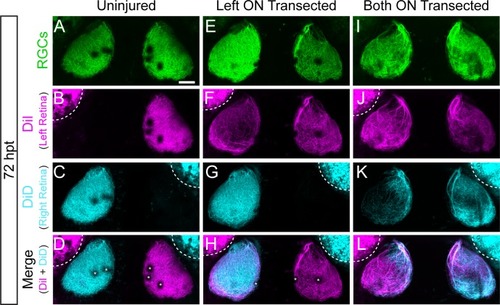
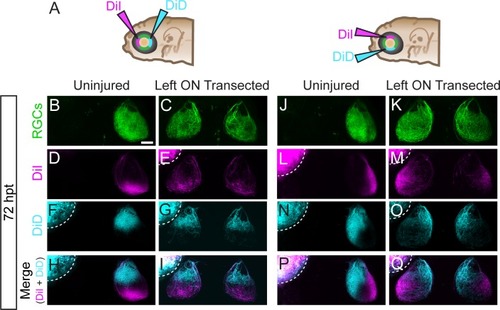
Transected RGC axons grow to ipsilateral and contralateral tecta. (A-L) Examples of RGC axonal tracings of Tg(isl2b:GFP) larvae at 72 hpt that were uninjured (A-D), received only a left optic nerve (ON) transection (E-H) or had both ON transected (I-L). Axons were traced by injecting the lipophilic dyes DiI and DiD into the left and right retina, respectively. RGC axons of uninjured nerves project to only contralateral tecta, while transected RGC axons grow to both ipsilateral and contralateral tecta. Asterisks indicate melanophores on the skin. Dashed lines outline dye fluorescence from the injected eye. Scale bar = 50 μm. |
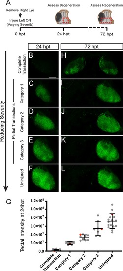
Partially injured RGC axons grow predominantly to contralateral tecta. (A) At 0 dpt, Tg(isl2b:GFP) larvae with the right eye removed received optic nerve transections ranging in severity from completely transected (n = 17 larvae), partially transected (Category 1, n = 8; Category 2, n = 9; Category 3, n = 8) to uninjured (n = 16). The extent of the transections was assessed at 24 hpt and regeneration to the tecta was examined at 72 hpt. Red dashed boxes indicate the imaged tectal areas. (B-F) Decreasing the severity of the transection spares more intact axons in the tectum at 24 hpt as shown by the GFP expression of RGC axons. (G) Quantification of the intensity of GFP signal in the right tectum at 24hpt. Red data points indicate the larvae represented in (B-F) images. (H-L) Reducing the severity of the injury reduces inappropriate growth to the ipsilateral tectum. Scale bar = 50 μm. |
Regenerating RGC axons terminate in correct topographic regions. (A) At 72 hpt, small populations of RGCs were labeled by DiI and DiD injections into anterior and posterior, or dorsal and ventral quadrants of the left retinas of Tg(isl2b:GFP)that were uninjured or received left optic nerve (ON) transection. The right eye was removed to facilitate analysis. (B-Q) Following optic nerve transection, RGC axons project to both the ‘incorrect’ ipsilateral and contralateral tecta yet maintain the same correct topographic specificity as uninjured RGCs; (B-I) anterior RGCs to posterior tectum and posterior RGCs to anterior tectum (uninjured, n = 22 nerves from 22 larvae; left ON transected, n = 21 nerves from 21 larvae), as well as dorsal RGCs to ventral tectum and ventral RGCs to dorsal tectum (uninjured, n = 23 nerves from 23 larvae; left ON transected, n = 16 nerves from 16 larvae). Dashed lines outline fluorescence from the injected eye. Scale bar = 50 μm. |