
Fig 6
- ID
- ZDB-FIG-190723-482
- Publication
- Harvey et al., 2019 - Optic nerve regeneration in larval zebrafish exhibits spontaneous capacity for retinotopic but not tectum specific axon targeting
- Other Figures
- All Figure Page
- Back to All Figure Page
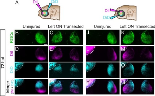
Regenerating RGC axons terminate in correct topographic regions. (A) At 72 hpt, small populations of RGCs were labeled by DiI and DiD injections into anterior and posterior, or dorsal and ventral quadrants of the left retinas of Tg(isl2b:GFP)that were uninjured or received left optic nerve (ON) transection. The right eye was removed to facilitate analysis. (B-Q) Following optic nerve transection, RGC axons project to both the ‘incorrect’ ipsilateral and contralateral tecta yet maintain the same correct topographic specificity as uninjured RGCs; (B-I) anterior RGCs to posterior tectum and posterior RGCs to anterior tectum (uninjured, n = 22 nerves from 22 larvae; left ON transected, n = 21 nerves from 21 larvae), as well as dorsal RGCs to ventral tectum and ventral RGCs to dorsal tectum (uninjured, n = 23 nerves from 23 larvae; left ON transected, n = 16 nerves from 16 larvae). Dashed lines outline fluorescence from the injected eye. Scale bar = 50 μm. |