
Fig 5
- ID
- ZDB-FIG-190723-481
- Publication
- Harvey et al., 2019 - Optic nerve regeneration in larval zebrafish exhibits spontaneous capacity for retinotopic but not tectum specific axon targeting
- Other Figures
- All Figure Page
- Back to All Figure Page
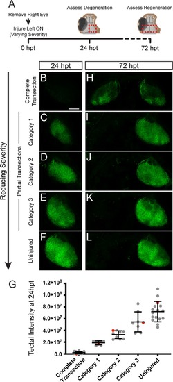
Partially injured RGC axons grow predominantly to contralateral tecta. (A) At 0 dpt, Tg(isl2b:GFP) larvae with the right eye removed received optic nerve transections ranging in severity from completely transected (n = 17 larvae), partially transected (Category 1, n = 8; Category 2, n = 9; Category 3, n = 8) to uninjured (n = 16). The extent of the transections was assessed at 24 hpt and regeneration to the tecta was examined at 72 hpt. Red dashed boxes indicate the imaged tectal areas. (B-F) Decreasing the severity of the transection spares more intact axons in the tectum at 24 hpt as shown by the GFP expression of RGC axons. (G) Quantification of the intensity of GFP signal in the right tectum at 24hpt. Red data points indicate the larvae represented in (B-F) images. (H-L) Reducing the severity of the injury reduces inappropriate growth to the ipsilateral tectum. Scale bar = 50 μm. |