
Fig 2
- ID
- ZDB-IMAGE-190723-521
- Publication
- Harvey et al., 2019 - Optic nerve regeneration in larval zebrafish exhibits spontaneous capacity for retinotopic but not tectum specific axon targeting
- All Figures
- Figures for Harvey et al., 2019
|
Fig 2
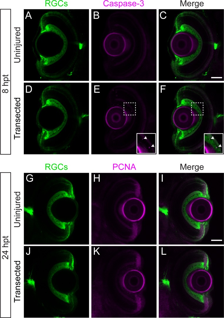
(A-F) Transverse optical sections of retinas from Tg(isl2b:GFP) uninjured larvae (A-C; n = 8 retinas) or larvae with transected optic nerves (D-E; n = 8 retinas) at 8 hpt labeled with anti-active caspase-3 (magenta). Following optic nerve injury, very few RGCs co-label with caspase-3 (E-F inset; see Table 1). (G-L) Transverse sections of retinas from Tg(isl2b:GFP) uninjured larvae (G-I; n = 10 retinas from 5 larvae) or larvae with transected optic nerves (J-L; n = 14 retinas from 7 larvae) at 24 hpt labeled with anti-PCNA (magenta) show no apparent PCNA staining in the inner nuclear layer. Scale bars = 50 μm.