
IMAGE
Fig 4
- ID
- ZDB-IMAGE-190723-524
- Publication
- Harvey et al., 2019 - Optic nerve regeneration in larval zebrafish exhibits spontaneous capacity for retinotopic but not tectum specific axon targeting
- All Figures
- Figures for Harvey et al., 2019
Image
|
Figure Caption
Fig 4
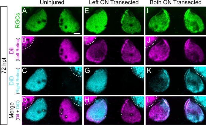
Transected RGC axons grow to ipsilateral and contralateral tecta.
(A-L) Examples of RGC axonal tracings of Tg(isl2b:GFP) larvae at 72 hpt that were uninjured (A-D), received only a left optic nerve (ON) transection (E-H) or had both ON transected (I-L). Axons were traced by injecting the lipophilic dyes DiI and DiD into the left and right retina, respectively. RGC axons of uninjured nerves project to only contralateral tecta, while transected RGC axons grow to both ipsilateral and contralateral tecta. Asterisks indicate melanophores on the skin. Dashed lines outline dye fluorescence from the injected eye. Scale bar = 50 μm.
Acknowledgments
This image is the copyrighted work of the attributed author or publisher, and
ZFIN has permission only to display this image to its users.
Additional permissions should be obtained from the applicable author or publisher of the image.
Full text @ PLoS One