
Fig 1
- ID
- ZDB-FIG-190723-477
- Publication
- Harvey et al., 2019 - Optic nerve regeneration in larval zebrafish exhibits spontaneous capacity for retinotopic but not tectum specific axon targeting
- Other Figures
- All Figure Page
- Back to All Figure Page
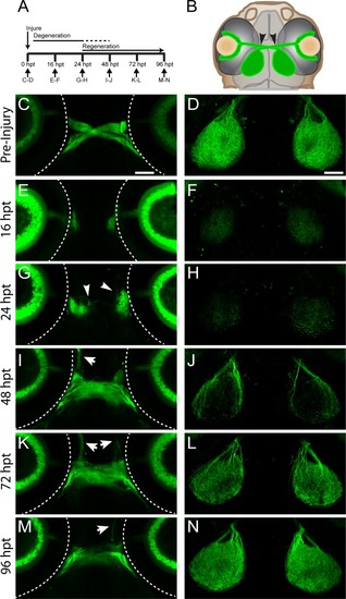
Regeneration of RGC axons in larval zebrafish. (A) Timeline of optic nerve regeneration. (B) Diagram of a Tg(isl2b:GFP) larva expressing GFP in RGCs and their axons. At 5dpf, optic nerves are transected with a sharpened tungsten needle proximal to the chiasm (black arrowheads). (C-D) Before injury, RGC axons cross the midline at the optic chiasm and innervate the contralateral tecta. Dashed lines indicate the outline of the eyes; scale bars = 50 μm. (E-F) Axonal growth from transected nerve at 16hpt is undetectable, while the portion of the nerve distal to the injury degenerates. (G-H) At 24 hpt, regrowing RGC axons begin to emerge from the proximal portion of the injured nerve (white arrowheads), while further degeneration continues. (I-J) By 48 hpt, regenerating axons project into the chiasm and re-innervate the tecta, though about 50% of transected nerves exhibit some misguided axonal growth (n = 81/160 nerves from 80 larvae, white arrows). (K-L) At 72 hpt, there is additional axonal growth to the tecta. (M-N) By 96 hpt, the axons within the chiasm fasciculate and robustly innervate the tecta (n = 160/160 nerves from 80 larvae). Representative images of chiasms and tecta for each timepoint are of the same fixed larva, though across timepoints are different larvae. |