- Title
-
Peli1b governs the brain patterning via ERK signaling pathways in zebrafish embryos
- Authors
- Kumar, A., Anuppalle, M., Maddirevula, S., Huh, T.L., Choe, J., Rhee, M.
- Source
- Full text @ Gene
|
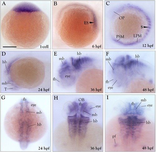
Expression of zebrafish peli1b is spatiotemporally restricted during embryonic development. (A–C) Lateral view of early stage embryos. (D–F) Lateral view and (G–I) dorsal view of late stage embryos. (A) Animal-pole view of an embryo showing that peli1b transcripts are abundant. (B) Animal-pole view of a shield stage embryo showing a greater level of peli1b transcripts on the dorsal side than on the ventral side. (C) At 12 hpf, there is peli1b expression in the hindbrain, somites, and presomitic mesodermregion. At 24 hpf, the lateral view (D) and dorsal view (G) show abundant peli1b expression in the telencephalon, diencephalon, and hindbrain (r 1-7), but a lack of expression in the midbrain–hindbrain boundary (MHB). At 36 hpf, the lateral view (E) and dorsal view (H) show abundant peli1b transcripts in the diencephalon, hindbrain, eyes, and pectoral fins. At 48 hpf, the lateral view (F) and dorsal view (I) show peli1b expression in the diencephalon, hindbrain (r 1-7), eyes, and pectoral fins. In the colorimetric experiments, samples were incubated for 3 h at room temperature. Abbreviations: ES, embryonic shield; OP, Optic premordium; LPM, lateral plate mesoderm; S, somites; PSM, pre-somitic mesoderm; hb, hindbrain; Mb, midbrain; T, telencephalon; fb, forebrain; OB, olfactory bulb. (n = 3). Scale bar- 50 μm. |
|
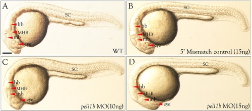
Morphological defects in zebrafish embryos after knockdown of peli1b. All images were taken at 24 hpf. (A) WT, (B) 5′ mismatch control, (C) peli1b MO (10 ng), and (D) peli1b MO (15 ng). Morpholino and 5′ mismatch control were injected at the 1-cell stage in synchronized embryos. (A–D) Lateral view of the embryos showing the phenotype after 10 ng or 15 ng injection of peli1b antisense nucleotidemorpholino. Abbreviations: NT, neural tube; SC, spinal cord; fb, forebrain; mb, midbrain; hb, hindbrain; MHB, midbrain–hindbrain boundary. (n = 3). Scale bar- 50 μm. PHENOTYPE:
|
|
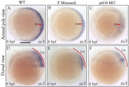
WISH distribution analysis of zic3 in peli1bmorphants at the shield stage. Animal pole view of zebrafish embryos at the shield stage (A–C). Dorsal view of zebrafish embryos at the shield stage (D–F). Images for WT (A & D), 5′ mismatch (B & E), and peli1b morphants injected with 10 ng morpholino at the 1-cell stage (C & F). Red arrowheads show zic3 expression and intensity in WT, 5′ mismatch, and peli1b morphants. The animal-pole view shows that the embryonic shield (EM) area has lower expression of zic3 transcripts in the peli1b knockdown embryos than in WT embryos. The dorsal view shows the reduced expression of zic3 in peli1b MO embryos. (n = 3). Abbreviation: ES- embryonic shield, DA- dorsal area. Scale bar- 50 μm. (For interpretation of the references to color in this figure legend, the reader is referred to the web version of this article.) |
|
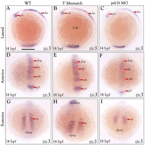
WISH distribution analysis of zic3 in peli1bknockdown embryos at 14 hpf. Lateral view of zebrafishembryos (A–C). Anterior view (D–F). Posterior view (G–I). WT embryos (A, D, & G), 5′ mismatch controls (B, E, & H), and peli1b morphants injected with 10 ng morpholino at the 1-cell stage (C, F, & I). Red arrowheads show zic3expression and intensity in WT, 5′ mismatch, and peli1bmorphants. (n = 3). Abbreviations: pd- posterior diencephalon, dsc- dorsal spinal cord, dnp- dorsal neural plate, hb- hindbrain, mb- midbrain, dpnp-dorsal posterior neural plate, sp-segmental plate. Scale bar- 50 μm. (For interpretation of the references to color in this figure legend, the reader is referred to the web version of this article.) EXPRESSION / LABELING:
PHENOTYPE:
|
|
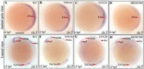
WISH distribution analysis of zic3 transcripts at the shield and 12 hpf stages in embryos incubated with ERK inhibitors. WT (A), vehicle control (B), UO126-treated (C), and SB203580-treated (D) embryos at the shield stage. WT (E), vehicle control (F), UO126-treated (G), and SB203580-treated (H) embryos at 12 hpf. WISH analysis of zic3 transcripts was performed after fixation. Shield-stage embryos are shown from the animal-pole view and 12 hpf embryos are shown from the dorsal view. Red arrowheads show the changes in zic3 expression in embryos treated with ERK inhibitors (SB203580 and UO126) compared with WT embryos. The embryonic shield region is shown in a shield-stage embryo with a red arrowhead. The posterior diencephalon, dorsal spinal cord, and tail bud are shown for the vehicle control in the 12 hpf embryos. (n = 3). Scale bar- 50 μm. (For interpretation of the references to color in this figure legend, the reader is referred to the web version of this article.) EXPRESSION / LABELING:
PHENOTYPE:
|

ZFIN is incorporating published figure images and captions as part of an ongoing project. Figures from some publications have not yet been curated, or are not available for display because of copyright restrictions. |

ZFIN is incorporating published figure images and captions as part of an ongoing project. Figures from some publications have not yet been curated, or are not available for display because of copyright restrictions. PHENOTYPE:
|

ZFIN is incorporating published figure images and captions as part of an ongoing project. Figures from some publications have not yet been curated, or are not available for display because of copyright restrictions. |

ZFIN is incorporating published figure images and captions as part of an ongoing project. Figures from some publications have not yet been curated, or are not available for display because of copyright restrictions. EXPRESSION / LABELING:
|
Reprinted from Gene, 694, Kumar, A., Anuppalle, M., Maddirevula, S., Huh, T.L., Choe, J., Rhee, M., Peli1b governs the brain patterning via ERK signaling pathways in zebrafish embryos, 1-6, Copyright (2019) with permission from Elsevier. Full text @ Gene

