
- Title
-
Microtubule asters anchored by FSD1 control axoneme assembly and ciliogenesis
- Authors
- Tu, H.Q., Qin, X.H., Liu, Z.B., Song, Z.Q., Hu, H.B., Zhang, Y.C., Chang, Y., Wu, M., Huang, Y., Bai, Y.F., Wang, G., Han, Q.Y., Li, A.L., Zhou, T., Liu, F., Zhang, X.M., Li, H.Y.
- Source
- Full text @ Nat. Commun.

FSD1 is required for ciliogenesis in human cells and zebrafish. a RPE-1 cells were transfected with the indicated siRNA and serum-starved for 48 h. Cells were stained with the indicated antibodies and examined by immunofluorescence microscopy. Scale bar, 5 μm. bEffects of FSD1 depletion on cilia formation in cycling (+Serum) or quiescent (−Serum) RPE-1 cells. Acetylated α-tubulin (Ac-tubulin) is ciliary marker. c Cilia defects induced by FSD1 knockdown in RPE-1 cells were rescued by expressing a GFP-tagged, RNAi-resistant form of FSD1. Data are presented as mean ± s.d. of three independent experiments. n number of cells. d, e Fsd1 morphants (aMO and sMO) displayed curved body and pericardial edema at 72 hpf. The arrows mark curved body and arrowheads mark pericardial edema. The fsd1-misMO were used as control. Scale bars, 1 mm. Data are presented as the mean ± s.d. of three independent experiments. f, g Fsd1 MOs (aMO and sMO) caused left-right asymmetry defects. The spaw probe was used to label the left lateral plate mesoderm in the whole-mount in situ hybridization at 18-somite stage. Scale bar, 150 μm. n number of fishes. h–jFsd1 MOs (aMO and sMO) impaired ciliogenesis in Kupffer’s vesicle at 10-somite stage (10 s). Bars indicate the median. Scale bar, 10 μm. k Fsd1 knockout impaired ciliogenesis in Kupffer’s vesicle at 10 s. Bars indicate the median. Scale bar, 10 μm. In all panels, statistical comparisons between two groups were carried out by two-tailed t-test. *P < 0.05, **P < 0.01, ***P < 0.001 EXPRESSION / LABELING:
PHENOTYPE:
|

(a) Fsd1 atg-morpholino efficiency was validated by western blot. (b) Fsd1 splice-morpholino efficiency was validated by RT-PCR and sequencing. The PCR product was 292 bp in control embryos and 194 bp in fsd1 sMO injected embryos. The sequencing result of the PCR product below showed deletion of exon 2 (98 bp) in fsd1 sMO injected embryos. Purple, orange and blue colors mark exon 1, 2 and 3, respectively. The red box indicates the fsd1 sMO targeted sequence. (c) Fsd1 MOs caused left-right asymmetry defects. The cmlc probe was used to label the heart tube in the whole-mount in situ hybridization at 26 hpf. Scale bar, 100 μm. n, number of fishes. (d) The target sequence of fsd1 aMO and sequence of aMO-resistant form of zebrafish fsd1 mRNA (fsd1 re-zmRNA). (e, f) Zebrafish fsd1 mRNA partially rescued the LR asymmetry defects induced by fsd1 aMO marked by spaw or cmlc. (g) Schematic diagram illustrates the difference between fsd1 re-zmRNA and fsd1-CC re-zmRNA. (h, i) Zebrafish fsd1-CC re-zmRNA could not rescue the LR asymmetry defects induced by fsd1 aMO marked by spaw or cmlc. Data are presented as mean ± s.d. of at least three independent experiments. NS, not significant, *P < 0.05, **P < 0.01, ***P < 0.001. n, number of fishes. |
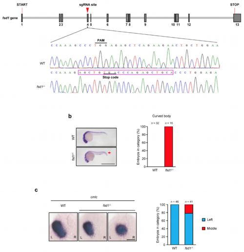
(a) Fsd1 knock out efficiency was validated by sequencing. (b) Fsd1 knock out displayed curved body at 72 hpf. The arrow marked curved body. Scale bars, 1 mm. (c) Fsd1 knock out caused left-right asymmetry defects. The cmlc probe was used to label the heart tube in the whole-mount in situ hybridization at 26 hpf. Scale bar, 100 μm. n, number of fishes. EXPRESSION / LABELING:
PHENOTYPE:
|