
Fig. 1
- ID
- ZDB-IMAGE-190701-31
- Genes
- Publication
- Tu et al., 2018 - Microtubule asters anchored by FSD1 control axoneme assembly and ciliogenesis
- All Figures
- Figures for Tu et al., 2018
|
Fig. 1
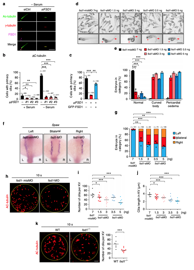
FSD1 is required for ciliogenesis in human cells and zebrafish. a RPE-1 cells were transfected with the indicated siRNA and serum-starved for 48 h. Cells were stained with the indicated antibodies and examined by immunofluorescence microscopy. Scale bar, 5 μm. bEffects of FSD1 depletion on cilia formation in cycling (+Serum) or quiescent (−Serum) RPE-1 cells. Acetylated α-tubulin (Ac-tubulin) is ciliary marker. c Cilia defects induced by FSD1 knockdown in RPE-1 cells were rescued by expressing a GFP-tagged, RNAi-resistant form of FSD1. Data are presented as mean ± s.d. of three independent experiments. n number of cells. d, e Fsd1 morphants (aMO and sMO) displayed curved body and pericardial edema at 72 hpf. The arrows mark curved body and arrowheads mark pericardial edema. The fsd1-misMO were used as control. Scale bars, 1 mm. Data are presented as the mean ± s.d. of three independent experiments. f, g Fsd1 MOs (aMO and sMO) caused left-right asymmetry defects. The spaw probe was used to label the left lateral plate mesoderm in the whole-mount in situ hybridization at 18-somite stage. Scale bar, 150 μm. n number of fishes. h–jFsd1 MOs (aMO and sMO) impaired ciliogenesis in Kupffer’s vesicle at 10-somite stage (10 s). Bars indicate the median. Scale bar, 10 μm. k Fsd1 knockout impaired ciliogenesis in Kupffer’s vesicle at 10 s. Bars indicate the median. Scale bar, 10 μm. In all panels, statistical comparisons between two groups were carried out by two-tailed t-test. *P < 0.05, **P < 0.01, ***P < 0.001