- Title
-
rbpms2 functions in Balbiani body architecture and ovary fate
- Authors
- Kaufman, O.H., Lee, K., Martin, M., Rothhämel, S., Marlow, F.L.
- Source
- Full text @ PLoS Genet.
|
Expression of rbpms2 in oocytes. Fluorescent in situ hybridization displaying Bb localization (arrow) of rbpms2a transcripts (A), while rbpms2b transcripts are not Bb-localized (C). Sense control probes demonstrate that rbpms2a and rbpms2b signals are specific (B,D). (E) RT-PCR on FACS-sorted granulosa and theca cells demonstrates that expression of rbpm2a and rbpms2b is germ cell specific. Antibody staining for Rbpms2 in ovaries mutant for rbpm2a (F) or rbpms2b (I) suggests both ohnologs are Bb localized where Bucky ball protein also resides (G,H,J,K). EXPRESSION / LABELING:
|
|
Rbpms2 embryonic phenotype characterization. (A) rbpms2a and rbpms2b alignment showing extremely similar exon structure, with the pink arrowheads pointing to the Crispr-Cas9 disrupted region of exon 5 in both rbpms2a and rbpms2b, and the blue arrowhead pointing to the disrupted splice site in rbpm2b. (B) Table of germ-line rbpms2 alleles and predicted protein structure, with single and compound mutant phenotypes represented in the two right columns. In the table, n = number of embryos of that genotype, and the number in parentheses represents independent parental crosses from which those embryos were derived. Representative images of the lateral view of d3 embryos, showing head and cardiac structures of non-mutant sibling (C) and rbpms2aae30/ae30; rbpms2bsa9329/sa9329 double mutant (D). Representative image of lateral view of d5 embryos, showing head and cardiac structures of non-mutant sibling (E) and recovered (F) and edematous (G) rbpms2aae30/ae30; rbpms2bsa9329/sa9329 double mutants. PHENOTYPE:
|
|
rbpms2 mutant allele stability and localization activity in somatic cells. (A) RT-PCR to detect rbpms2a and rbpms2b maternal transcripts. Pink asterisks indicate likely nonsense mediated decay of mutant allele transcripts. (B-F) Wild-type GFP-Rbpms2a/b (B, D) and GFP-Rbpms2bsa9329 (F) localize to granules in HEK 293 cells, while GFP-Rbpms2aae30 and GFP-Rbpms2bae32 are not localized. (G-K) Zebrafish blastula cells expressing GFP-Rbpms2 fusions. (G,H) Wild-type GFP-Rbpms2b localizes near the nucleus (H), is apparently associated with the centrosome/spindle in some cells, and (G) is in granules that are not positive for the stress granule marker Tial-1. (H) A subset of GFP-Rbpms2b positive granules are positive for the p-body marker Dcp2 (open arrowheads). (I) GFP-Rbpms2bsa9329 localization to the centrosome/spindle but not granules. (J) GFP-Rbpms2bae32 and (K) GFP-Rbpms2aae30 localization. Insets show magnified views of the highlighted cells. Images are representative slices from Z-stacks of sphere stage embryos viewed from the animal pole. |

ZFIN is incorporating published figure images and captions as part of an ongoing project. Figures from some publications have not yet been curated, or are not available for display because of copyright restrictions. EXPRESSION / LABELING:
|
|
Zebrafish rbpms2 mutants are fertile males. (A-B) Non-mutant siblings of rbpms2 double mutants can differentiate and mature as fertile females with overtly normal oocytes observed when germ cells are marked by ziwi:GFP (A) or with H&E staining (B). (C-D) Non-mutant male siblings of rbpms2 double mutants display normal spermatogenesis with spermatogonia (SG) and spermatozoa (SZ) detectable with ziwi:GFP (C) or with H&E staining (D). (E-F) rbpms2aae30;rbpms2bsa9329 double mutant males have no distinguishable differences in testis morphology or spermatogenic stages from their non-mutant male siblings. (G) Stacked bar chart depicts male to female ratio of adults recovered from each rbpms2a;rbpms2b genotype (blue = male, pink = female). |
|
rbpms2 is required for definitive female differentiation. (A) Quantification of PGC numbers in d3 rbpms2 double mutants and non-mutant siblings. (B-C) Vasa staining to count PGCs in a representative slice from Z-series with PGCs circled in yellow in non-mutant sibling (B), and rbpms2 double mutant (C). (D-E) Transgenic germ cell marker ziwi:GFP allows visualization of comparable gonocyte development in bipotential gonads of d21 non-mutant siblings (D) and rbpms2 mutants (E). (F-G) Female (F) and male (G) gonad differentiation of control non-mutant siblings at d35. (H) rbpms2 mutant gonad with oocyte-like cells outlined in solid line, and testis-like cells outlined in dashed line. (I) rbpms2 mutant gonad resembling non-mutant male siblings. EXPRESSION / LABELING:
PHENOTYPE:
|
|
rbpms2 mutant oocytes contain atypical cytoplasmic inclusions. (A-B) Comparable clusters of gonocytes from d21 non-mutant siblings (A), and rbpms2 double mutants (B). (C,D) Oocytes from d35 non-mutant siblings (C), and rbpms2 double mutants (D). Marked features include: n = nucleoli, white dotted line outlining areas of mitochondrial enrichment, yellow arrows indicating nuage accumulations, pink arrows indicating atypical cytoplasmic inclusions. Areas marked in a black box in (C, D) are magnified in (C’,D’). (C’, D’) High magnification images of nuage with associated mitochondria (white arrows) in non-mutant oocyte (C’), and juxtaposed nuage and atypical cytoplasmic inclusion in rbpms2 mutant oocyte (D’). (E-G) Oocytes of comparable diameter (E) were used to quantify and compare number of nuage aggregates over 0.5μm (F), and number of cytoplasmic inclusions figover 1μm (G). Significance was analyzed with unpaired t-test. PHENOTYPE:
|
|
Bucky ball stained structures are abnormal in rbpms2 mutants. (A) Non-mutant sibling males have no specific Buc signal. (B) Non-mutant sibling female with asymmetric Buc localization in compact perinuclear domain of primary (stage I) oocytes (arrows). (B’) Magnified view of normal Buc localization in non-mutant sibling oocyte. (C) Like their control male siblings, differentiated male rbpms2 mutants have no Buc signal. (D) Oocyte-like cells of rbpms2 mutant gonads have more dispersed asymmetric Buc staining, frequently observed in a ring-like pattern (arrow-heads). (D’) Magnified view of ring-like Buc localization in rbpms2 mutant oocyte. EXPRESSION / LABELING:
|
|
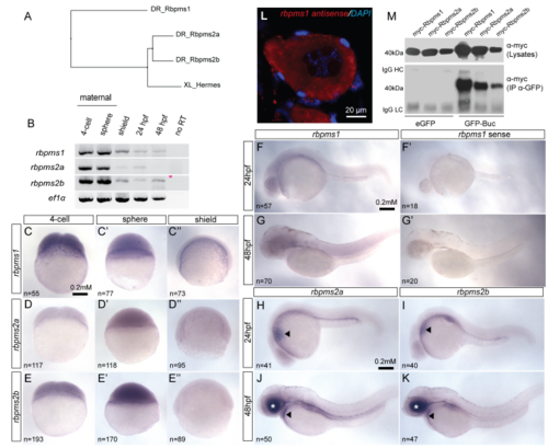
Conserved expression of Xhermes homologs in zebrafish. (A) Phylogenetic tree clustered according to protein sequence similarity between Xenopus Hermes and zebrafish Hermes homologs Rbpms1, Rbpms2a, and Rbpms2b. (B) RT-PCR on maternal and zygotic stages of zebrafish embryonic development demonstrate all three RNAs are abundant as maternal transcripts. (CE’’) Lateral views of whole-mount in situ hybridization (ISH) for rbpms1, rbpms2a and rbpms2b in 4-cell, sphere, and shield- stage embryos. Lateral views of 24 hpf ISH for rbpms1 (F), and sense probe control (F’). Lateral views of 48 hpf ISH for rbpms1 (G), and sense probe control (G’). Lateral views of 24 hpf embryos shows similar rbpms2a (H) and rbpms2b (I) ISH staining in heart primordium (arrow heads). Lateral view of 48 hpf embryos reveals similar rbpms2a (J) and rbpms2b (K) ISH staining pattern in the heart (arrow head) and retina (white asterisk). (L) Fluorescent in situ hybridization displaying unlocalized rbpms1 transcripts in primary zebrafish oocyte. (M) GFP immunoprecipitation of Myc- tagged Rbpms1, Rbpms2a and Rbpms2b demonstrates that that all three proteins can interact with GFP-Bucky ball in vitro. |
|
Rbpms2, Vasa and Macf1/Mgn in rbpms2 mutant ovaries. Antibody staining for Rbpms2 (magenta), Vasa (green), MacF1 (red) and DAPI (blue) proteins in juvenile ovaries (d35) of (A) single mutant for rbpm2a (B) single mutant for rbpm2b, and (C) double mutant. (D-F) rRbpms2 antibody staining (red) and DAPI (blue) in adult ovary of (D) WT, (E) single mutant for rbpm2a (F) single mutant for rbpm2b. n=5 gonads per genotype. |
|
RNA transcripts in rbpms2 transgenic ovaries. Whole-mount in situ hybridization on ovary fragments was performed using anti-sense apple (RFP) probe and colorimetric probe detection. (A) Control transgenic negative (wild-type) ovaries demonstrate the baseline staining for this probe. (B,C) Ovaries from Tg[buc:RFP-rbpms2b] and Tg[buc:RFP-rbpms2bΔRNP1] both display alkaline phosphatase staining, indicating the presence of apple-rbpms2b (B) and apple-rbpms2bΔRNP1 (C) transcripts in oocytes. |
|
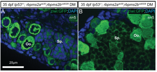
Tp53 mutation does not modify rbpms2 DM phenotype. Transgenic germ cell marker ziwi:GFP allows visualization of comparable intersex germ cell phenotype in d35 gonads of tp53 M214K/+ ;rbpms2DM (A) and tp53 M214K/ M214K;rbpms2DM (B). Oo. = oocyte-like cell; Sp.= spermatocyte-like cells. |


