
- Title
-
Evidences for a New Role of miR-214 in Chondrogenesis
- Authors
- Roberto, V.P., Gavaia, P., Nunes, M.J., Rodrigues, E., Cancela, M.L., Tiago, D.M.
- Source
- Full text @ Sci. Rep.
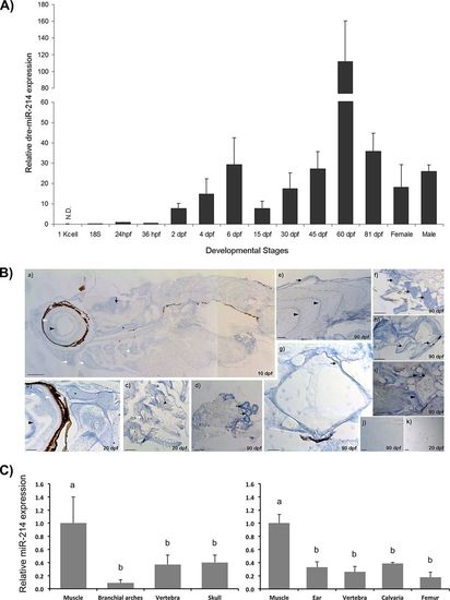
miR-214 expression correlates with skeletal elements in zebrafish. (A) Expression of miR-214 during zebrafish development, determined by miRNA qPCR. Values were normalized using zebrafish U6 small RNA and 24 hpf as reference sample and represent the mean ± s.d. of at least 3 independent replicates. hpf hours post fertilization, dpf days post fertilization, N.D. non-detected. Gap in the y-axis separates two different scales. (B) Detection of miR-214 by in situ hybridization in zebrafish with 10 (a), 20 (b, c, d) and 90 (e, f, g, h, i) dpf. From head to tail, miR-214 was detected in eye lens (arrowhead, a, b), retina (white arrowheads, a, b), brain (arrow, a), chondrocranium (asterisk, a, b), pharyngeal cartilage (white arrows, a, c), kidney (arrows, d), scales (arrow, e), muscle myotomes (arrowheads, e), cartilage in the base of pectoral fins (arrows, f), notochordal sheath (arrow, g), osteoid of haemal arches (arrows, h) and growth zones of vertebral body (arrowheads, i). Hybridization with negative control (scrambled) probe did not produce detectable signal, as observed in 90 and 20 dpf specimens (j and k, respectively). Scale bars: 0.2 mm for a, e, f and k; 0.1 mm for b, c, d, h, i and j; and 0.05 mm for g. (C) Relative expression of miR-214 in zebrafish (left panel) and mouse (right panel) adult tissues, determined by miRNA qPCR. Values were normalized using U6 small RNA and muscle as reference sample and represent the mean ± s.d. of at least 3 independent replicates (one-way Anova, different letters indicate statistical significance, p < 0.05). B. arches, Branchial arches. dpf days post fertilization. |
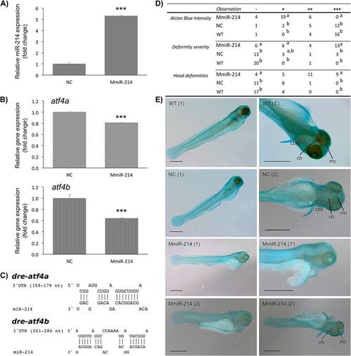
miR-214 ectopic expression downregulates atf4 transcripts and alters cranial cartilages of zebrafish. Larvae were microinjected at 1-cell stage with MmiR-214 or NC (18 μM) and analysed at 3 dpf. (A) Expression of miR-214 was determined by miRNA qPCR and normalized using U6 small RNA. (B) Expression of atf4a and atf4b in zebrafish larvae microinjected with MmiR-214 or NC. Levels of atf4a and atf4b transcripts expressions were determined by qPCR and normalized using 18 S ribosomal RNA housekeeping gene (similar results were obtained using ef1α housekeeping gene; data not shown). Results are presented as fold change over NC. Asterisks indicate values statistically different from NC (data are the mean ± s.d. of at least 3 independent replicates; Student’s t-test, ***p < 0.001). (C) Predicted miR-214 binding sites in zebrafish atf4a and atf4b 3′ UTRs. Zebrafish atf4a and atf4b transcripts sequences were collected from NCBI database and analysed using RNAhybrid. (D) Morphological alterations observed on 3 dpf embryos injected with MmiR-214 (n = 29) or NC (n = 21), and wild type (WT; n = 21), stained with alcian blue. Alcian blue intensity was classified as very weak (−), weak (+), normal (++) or intense (+++), while deformities present in the embryos were classified as absent/normal fish (−), low severity (+), severe (++), extremely severe (+++). Different letters indicate statistically significant differences between MmiR-214, NC and WT within the same classification (Chi-square test, p < 0.05). (E) Phenotype alterations observed in miR-214-injected embryos comparing to WT and NC-injected embryos, stained with alcian blue. Arrowheads indicate head deformations resulting from flattening of the mandible; arrows indicate differences on staining intensities of main cartilaginous structures (either absent or weak in miR-214-injected embryos). cbs, cerathobranchial cartilages; ch, ceratohyal; mc, Meckel’s cartilage. (1) or (2) indicate different embryos; (‘) indicate magnifications of the same embryo. Scale bar is 0.5 mm. EXPRESSION / LABELING:
|

miR-214 ectopic expression downregulates chondrogenic markers and impairs cranial cartilage formation in zebrafish. (A) Expression of marker genes of chondrogenesis. Expression levels of sox9a, sox9b, sox10, col2a1a, col10a1a, runx2a and mgp were evaluated by standard qPCR of 3 dpf zebrafish embryos microinjected with MmiR-214 or NC (18 μM), and normalized using 18 S ribosomal RNA housekeeping gene (similar results were obtained using ef1α housekeeping gene; data not shown). Results are presented as fold change over NC. Asterisks indicate values statistically different from NC (data are the mean ± s.d. of at least 3 independent replicates; one-way Anova, ***p < 0.001; **p < 0.01, *p < 0.05). (B) Histological characterization of miR-214 overexpression on zebrafish cartilage formation. 3 dpf embryos microinjected with NC (a) or MmiR-214 (b) were embedded in paraffin, sectioned and stained with the safranin-O/fast green/Mayer’s haematoxylin. Zebrafish eyes (e) are generally underdeveloped in miR-214-injected embryos when compared to NC. Cartilage associated proteoglycans (evidenced by safranin-O) are either absent or present in low amounts in the Ethmoid plate (Ep) or hyosympletic (Hs), respectively, of miR-214-injected embryos comparing to NC. Sections are in the coronal plane; (‘) and (‘’) indicate magnifications of the same section. EXPRESSION / LABELING:
|
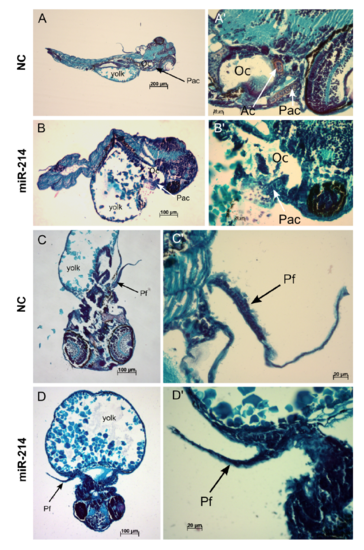
Histological characterization of miR-214 effect in zebrafish. Zebrafish embryos (3 dpf) injected with NC (A and C) or miR-214 mimic (B and D) were embedded in paraffin, sectioned and stained with the Safranin-O/Fast Green/Mayer’s Haematoxylin staining. In NC embryos, pharyngeal arch cartilage (Pac) (arrow in A and A’) and chondrocytes of the pectoral fins (Pf) (arrow in C and C’) are clearly stained with Safranin-O, but not in miR-214- injected embryos (arrows in B, B’, D and D’). Ac, auditory capsule; Pac, pharyngeal arch cartilage; Oc, otic capsule; Pf, pectoral fin; (‘) indicate magnification of the same section. |