
- Title
-
Extraocular Source of Oligodendrocytes Contribute to Retinal Myelination and Optokinetic Responses in Zebrafish
- Authors
- Tian, C., Zou, S., Hu, B.
- Source
- Full text @ Invest. Ophthalmol. Vis. Sci.
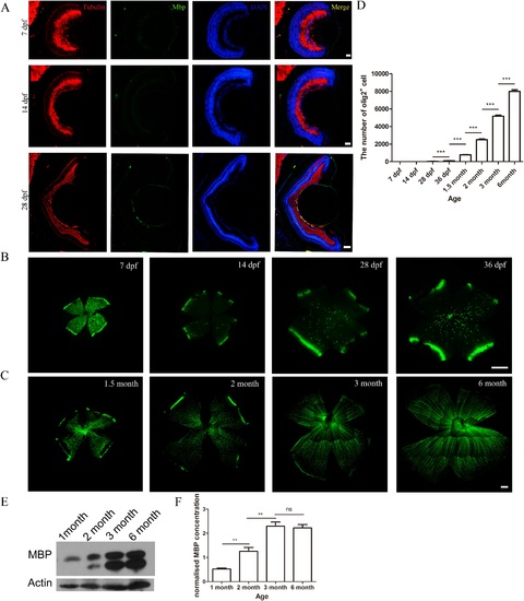
Development of myelination in zebrafish retinas. (A) Cryosections of 7- to 28-dpf zebrafish retinas were stained with MBP and a tubulin antibody. Myelin basic protein appeared at 28 dpf. (B, C) Whole-mount olig2 zebrafish retinas at the age of 20 to 36 dpf and 1.5 to 6.0 months. The olig2+ cells, outside the nerve fiber layer, were not OLs at 7 and 14 dpf. (D) Quantification of the number of OLs (n = 10, 10, 44, 21, 14, 13, 10, and 8, respectively) in zebrafish retinas from 7 dpf to 6 months. (E, F) Myelin basic protein Western blot of zebrafish retinas and its quantitative analysis at age 1.5 to 6.0 months. Scale bars: (A) = 100 µm; (B, C) = 200 µm. |
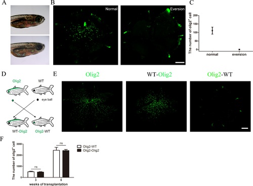
The origin of OLs in zebrafish retinas. (A) An illustrative picture shows the normal and eye eversion zebrafish. (B, C) Whole mount and quantification of the number of OLs in normal olig2 zebrafish retinas and the retinas after eye eversion 2 weeks later. (D) An illustrative picture shows the exchange of eyeballs in WT and olig2 zebrafish. (E) Whole-mount retinas show OLs in normal olig2 zebrafish eyes, olig2 zebrafish with WT eyes, and WT zebrafish with olig2 eyes at 3 weeks after eye eversion. (F) Quantification of the number of OLs in normal olig2 zebrafish eyes and olig2 zebrafish with WT eyes (n = 5 and n = 7, respectively) at 3 and 5 weeks after eye eversion. Scale bars: (B) = 150 µm, (E) = 200 µm. |
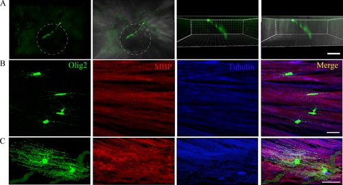
Oligodendrocytes at different developmental stages in zebrafish retinas. (A) The OPC was migrating out of the optic nerve head. The white dashed circles stand for optic nerve head. (B, C) Oligodendrocytes at different developmental stages: immature and mature OLs in zebrafish retinas stained with MBP and a tubulin antibody. The immature OLs are not colocalized with MBP, but the mature OLs are colocalized with MBP. Scale bars: 20 µm. |
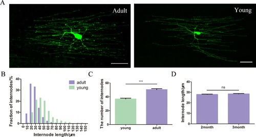
Oligodendrocytes at different stages have different behaviors. (A) The morphology of adult and young OLs in zebrafish retinas. (B) Distributions of internode lengths for adult OLs and young OLs (n = 2539 and n = 713 internodes, respectively). The K-S test shows that the internode length distribution was significantly different for young and adult OLs (P < 10-4). (C) Quantification of the number of internodes in adult and young OLs (n = 87 and n = 47, respectively). (D) Quantification of the lengths of internodes in adult OLs at 2 and 3 months after transplantation (n = 1354 and n = 1185, respectively). Scale bar: 20 µm. |

The relationship between myelination and the axon diameter. (A) Electron micrographs of RGC axons in zebrafish retinas from 1.5 to 3.0 months. (B, C) Quantification of the axon diameter (n = 517, 295, and 311 at 1.5, 2.0, and 3.0 months, respectively), myelinated (axons: n = 85, 140, and 251; zebrafish: n = 3, 3, and 3; images: n = 10, 11, and 13 at 1.5, 2.0, and 3.0 months), and unmyelinated axon (axons: n = 432, 155, and 60; zebrafish: n = 3, 3, and 4; images: n = 10, 11, and 16 at 1.5, 2.0, and 3.0 months) diameter in zebrafish retinas at the age of 1.5 to 3.0 months. (D) A scatterplot of axon diameter versus G-ratio for myelinated axons in 1.5-, 2.0-, and 3.0-month-old zebrafish retinas. (E) Quantification of the thickness of myelin in zebrafish retinas (axons: n = 82, 67, and 144; zebrafish: n = 3, 3, and 4; images: n = 10, 11, and 16 at 1.5, 2.0, and 3.0 months). Scale bar: 200 nm. |
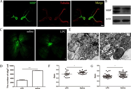
The OLs in zebrafish retinas are functional. (A) Cryosections of normal (right eye) and demyelinated (left eye) zebrafish retinas were immunolabeled with MBP and a tubulin antibody. (B) Myelin basic protein Western blot of normal and demyelinated zebrafish retinas. (C) Whole mount of normal and demyelinated olig2 zebrafish retinas. (D) Quantification of the number of OLs in the normal and demyelinated olig2 zebrafish retinas. (E) Electron micrographs of RGC axons in normal and LPC-induced demyelinated zebrafish retinas. (F, G) Optokinetic reflex behavior test of saline-treated and LPC-induced demyelinated zebrafish at the speed of 12 (n = 19, 18) and 24 rpm (n = 38, 41). Scale bars: (A) = 100 µm, (C) = 200 µm, and (E) = 200 nm. |