
FIGURE
Fig. 3
- ID
- ZDB-FIG-160517-46
- Publication
- Tian et al., 2016 - Extraocular Source of Oligodendrocytes Contribute to Retinal Myelination and Optokinetic Responses in Zebrafish
- Other Figures
- All Figure Page
- Back to All Figure Page
Fig. 3
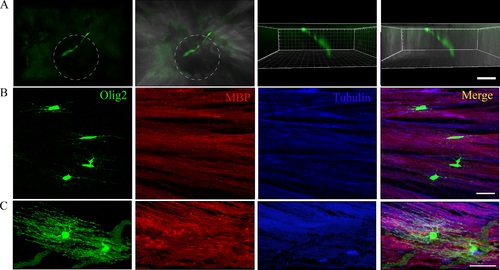
Oligodendrocytes at different developmental stages in zebrafish retinas. (A) The OPC was migrating out of the optic nerve head. The white dashed circles stand for optic nerve head. (B, C) Oligodendrocytes at different developmental stages: immature and mature OLs in zebrafish retinas stained with MBP and a tubulin antibody. The immature OLs are not colocalized with MBP, but the mature OLs are colocalized with MBP. Scale bars: 20 µm. |
Expression Data
Expression Detail
Antibody Labeling
Phenotype Data
Phenotype Detail
Acknowledgments
This image is the copyrighted work of the attributed author or publisher, and
ZFIN has permission only to display this image to its users.
Additional permissions should be obtained from the applicable author or publisher of the image.
Full text @ Invest. Ophthalmol. Vis. Sci.